
Bronchiole to respiratory bronchiole transition
This image shows the transitions that occur as a bronchiole becomes a terminal bronchiole which, in turn, becomes a respiratory bronchiole. Because alveoli are present in the wall of a respiratory bronchiole, this passageway is the first segment that serves a respiratory function. 40x

Bronchiole >
Cartilage and mixed glands have disappeared from the wall of a bronchiole. Large bronchioles are lined by pseudostratified columnar epithelium with cilia while the smallest (terminal) bronchioles have a simple columnar epithelium with cilia and club cells, but no goblet cells. Bronchioles are surrounded by alveoli, indicating they are intrapulmonary passages.

Terminal bronchiole
Cartilage and mixed glands have disappeared from the wall of a bronchiole. Large bronchioles are lined by pseudostratified columnar epithelium with cilia while the smallest (terminal) bronchioles have a simple columnar epithelium with cilia and club cells, but no goblet cells. Bronchioles are surrounded by alveoli, indicating they are intrapulmonary passages.

Respiratory bronchiole >
Respiratory bronchioles are smaller than bronchioles. Alveoli form an integral part of the wall of the respiratory bronchiole, indicating that respiratory bronchioles are capable of gas exchange and, therefore, are part of the respiratory portion of the respiratory system.

Alveolar ducts >
Alveoli in the walls of respiratory bronchioles gradually increase in number, reducing the surface area of the wall, thereby forming an alveolar duct, whose walls consist entirely of alveoli. The “wall” of an alveolar duct consists of connected rings that provide the framework for the openings of the attached alveoli. When cut in section, these rings look like “knobs” that support the alveoli. Alveolar ducts terminate in blind-ended alveolar sacs.
Alveoli >
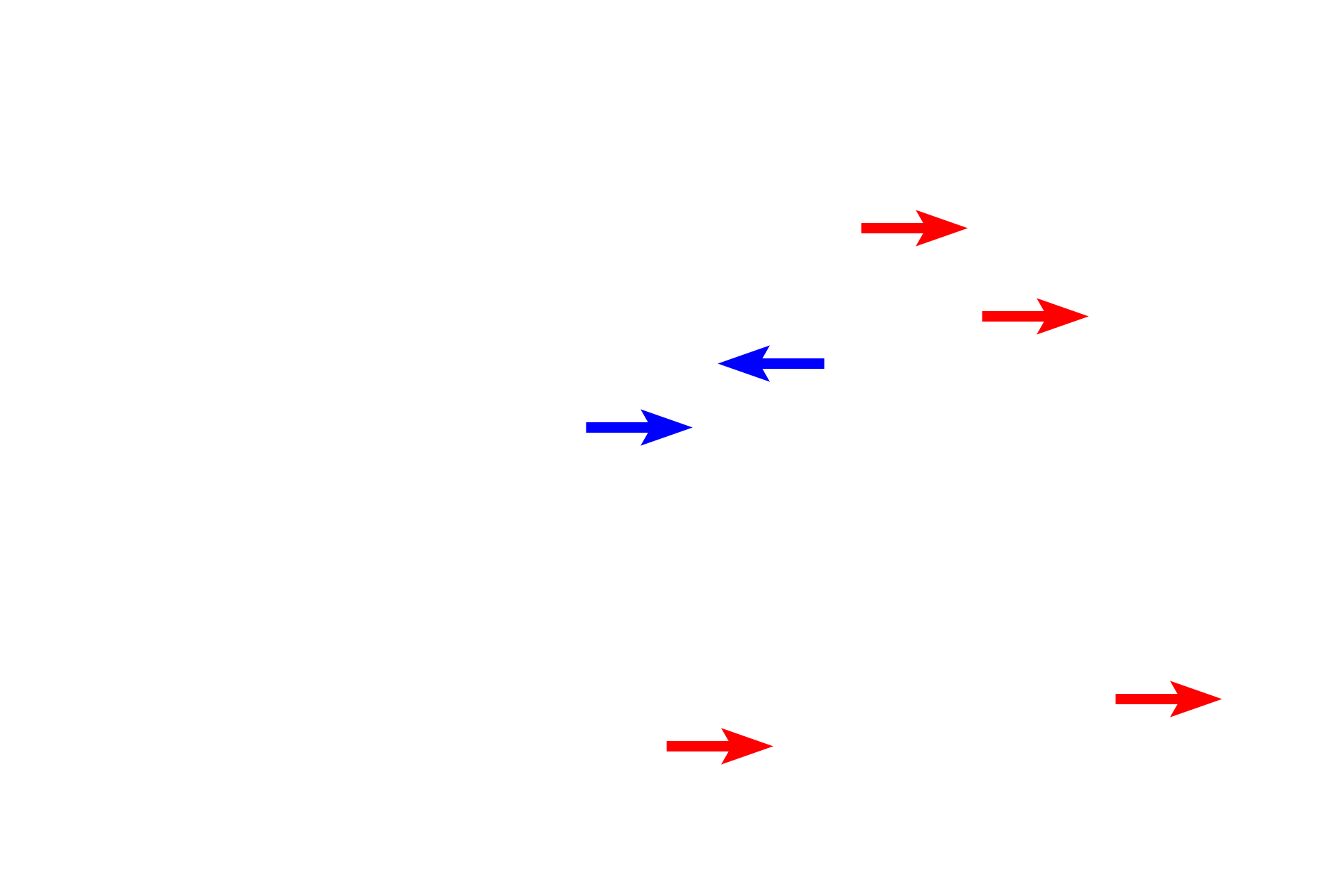
Alveoli are cup-shaped sacs, that first appear in the wall of respiratory bronchioles (blue arrows). They also form the majority of the wall of alveolar ducts and alveolar sacs (red arrows). The partition between adjacent alveoli, the, interalveolar septum, possesses numerous capillaries where gas exchange occurs.

Pulmonary vessels >
Pulmonary vessels consist of pulmonary arteries and veins. Pulmonary arteries, which deliver oxygen-poor blood to alveoli, travel adjacent to respiratory passageways. Pulmonary veins, which carry oxygen-rich blood from away alveoli, travel in the pleura or in intersegmental connective tissue independently of arteries. As veins near the root of the lung, they join the bronchi and pulmonary arteries.