
Trachea and primary bronchus
The walls of the trachea and primary bronchi are identical, resembling the pattern typical for most respiratory passageways. Longitudinally oriented elastic fibers are numerous in the lamina propria, sometimes forming an obvious elastic lamina. Bundles of interlacing smooth muscle lie external to the elastic fibers. The trachea and primary bronchi are extrapulmonary and are part of the conducting portion of the respiratory system. 100x
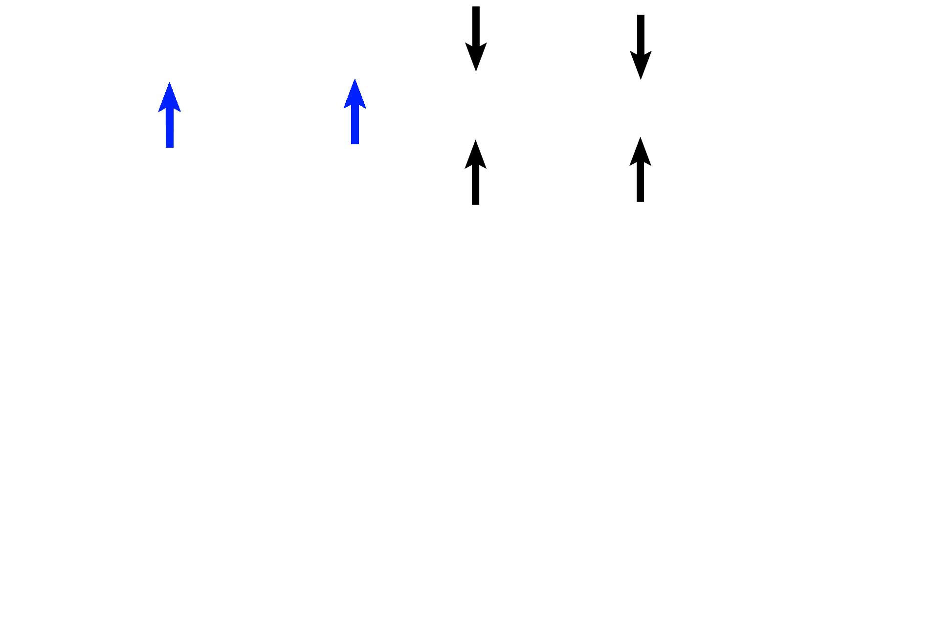
Epithelium >
Pseudostratified columnar epithelium (between black arrows) with cilia and goblet cells lines the trachea and primary bronchi. A prominent basement membrane (blue arrows) is typical for these two passageways.
Lamina propria >
A lamina propria extends from the basement membrane to the perichondrium. Elastic fibers form a distinct layer in this image, but the interlacing bundles of smooth muscle are difficult to identify
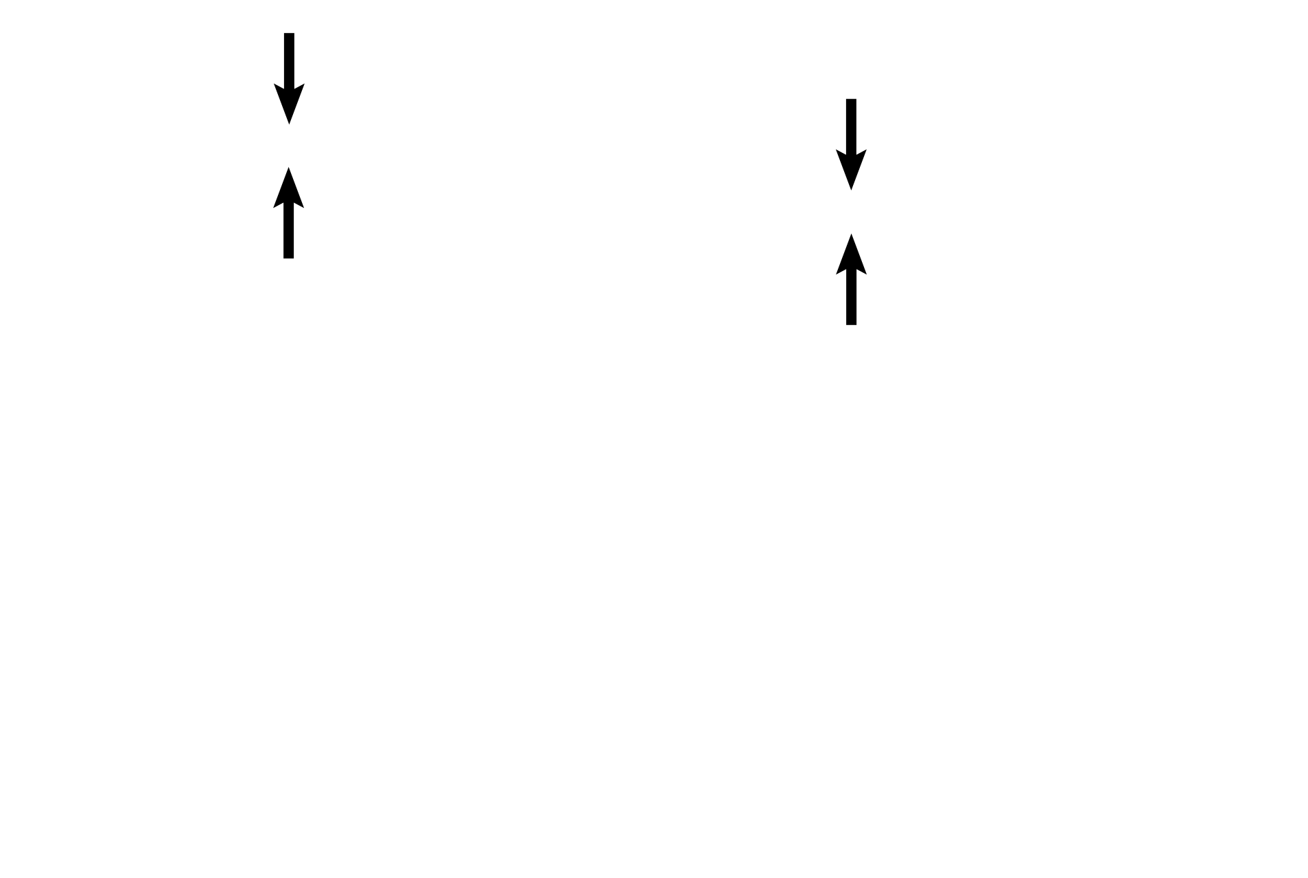
- Elastic lamina
A lamina propria extends from the basement membrane to the perichondrium. Elastic fibers form a distinct layer in this image, but the interlacing bundles of smooth muscle are difficult to identify
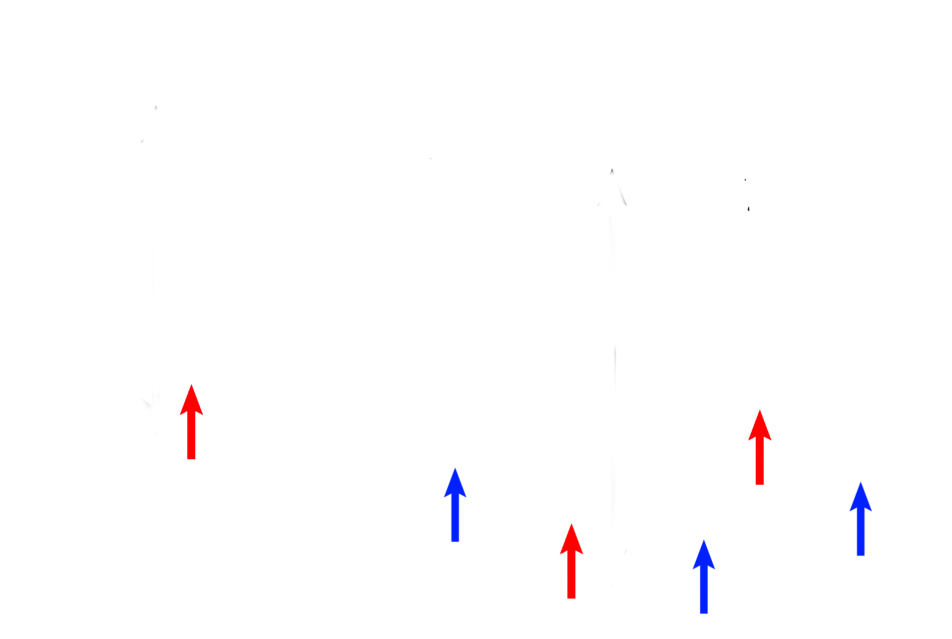
- Mixed glands >
Mixed glands (mucous cells, blue arrows; serous cells, red arrows) are also located in the lamina propria. Secretions from these glands provide a coating for these passsageways that entrap inhaled dust and other particulate matter.
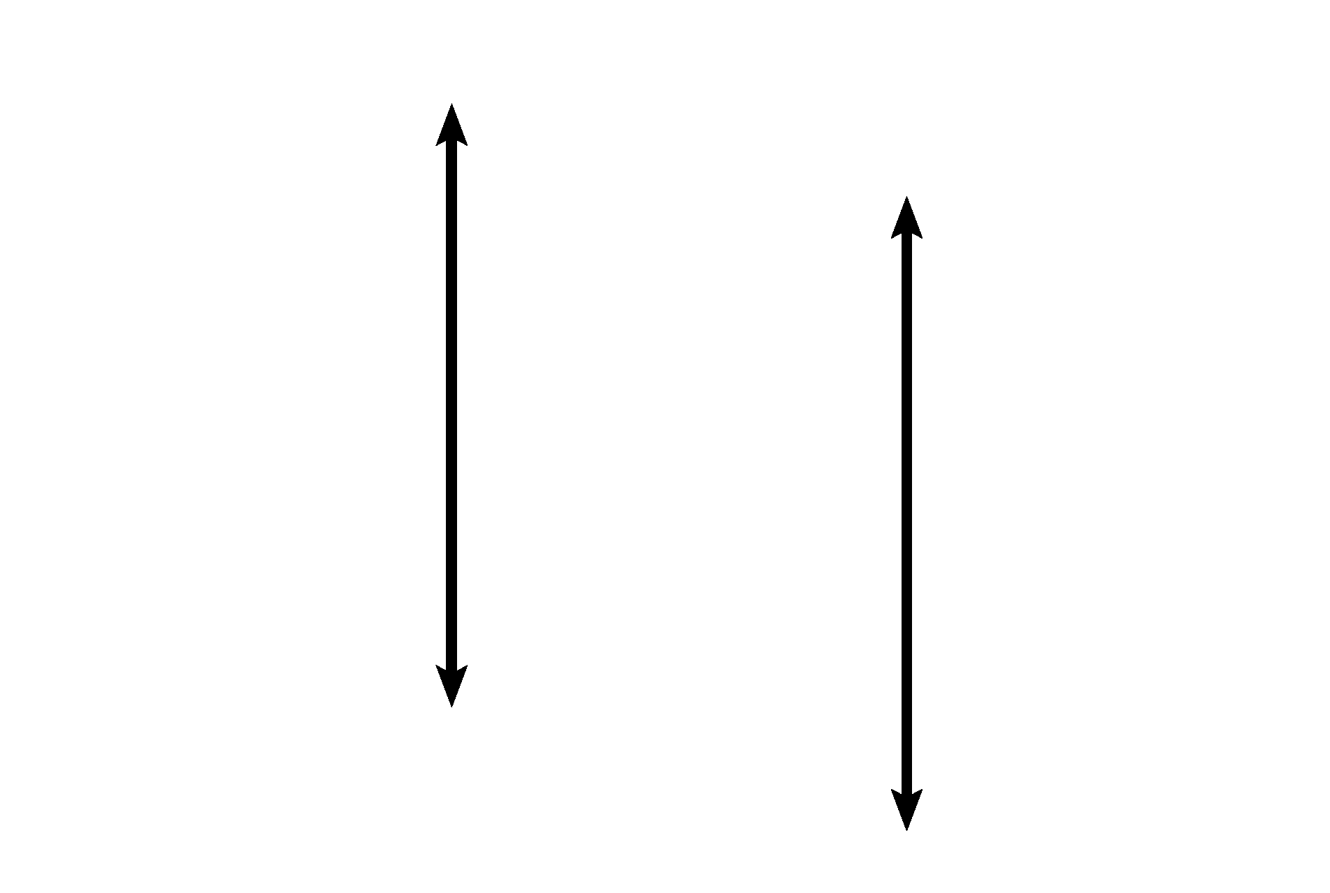
Cartilage >
Patency of the trachea and primary bronchi is maintained by a series of hyaline cartilage rings (black arrows). A perichondrium (blue arrows) surrounds this tissue.
Image source >
This image was taken from a slide in the University of Michigan slide collection.