Epididymis: body and tail
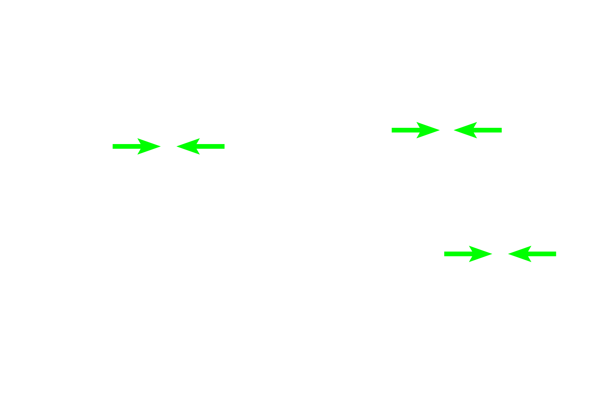
The body and tail of the epididymis are filled with the highly coiled duct of the epididymis. Numerous sectioned profiles of the single duct are seen in this image. The duct is lined by a very tall, regular, pseudostratified columnar epithelium with stereocilia, beneath which is layer of smooth muscle. As the duct progresses from the head to the tail region, its epithelium decreases in height and the smooth muscle layer increases in thickness. 40x

Duct of the epididymis
The body and tail of the epididymis are filled with the highly coiled duct of the epididymis. Numerous sectioned profiles of the single duct are seen in this image. The duct is lined by a very tall, regular, pseudostratified columnar epithelium with stereocilia, beneath which is layer of smooth muscle. As the duct progresses from the head to the tail region, its epithelium decreases in height and the smooth muscle layer increases in thickness. 40x

- Epithelium
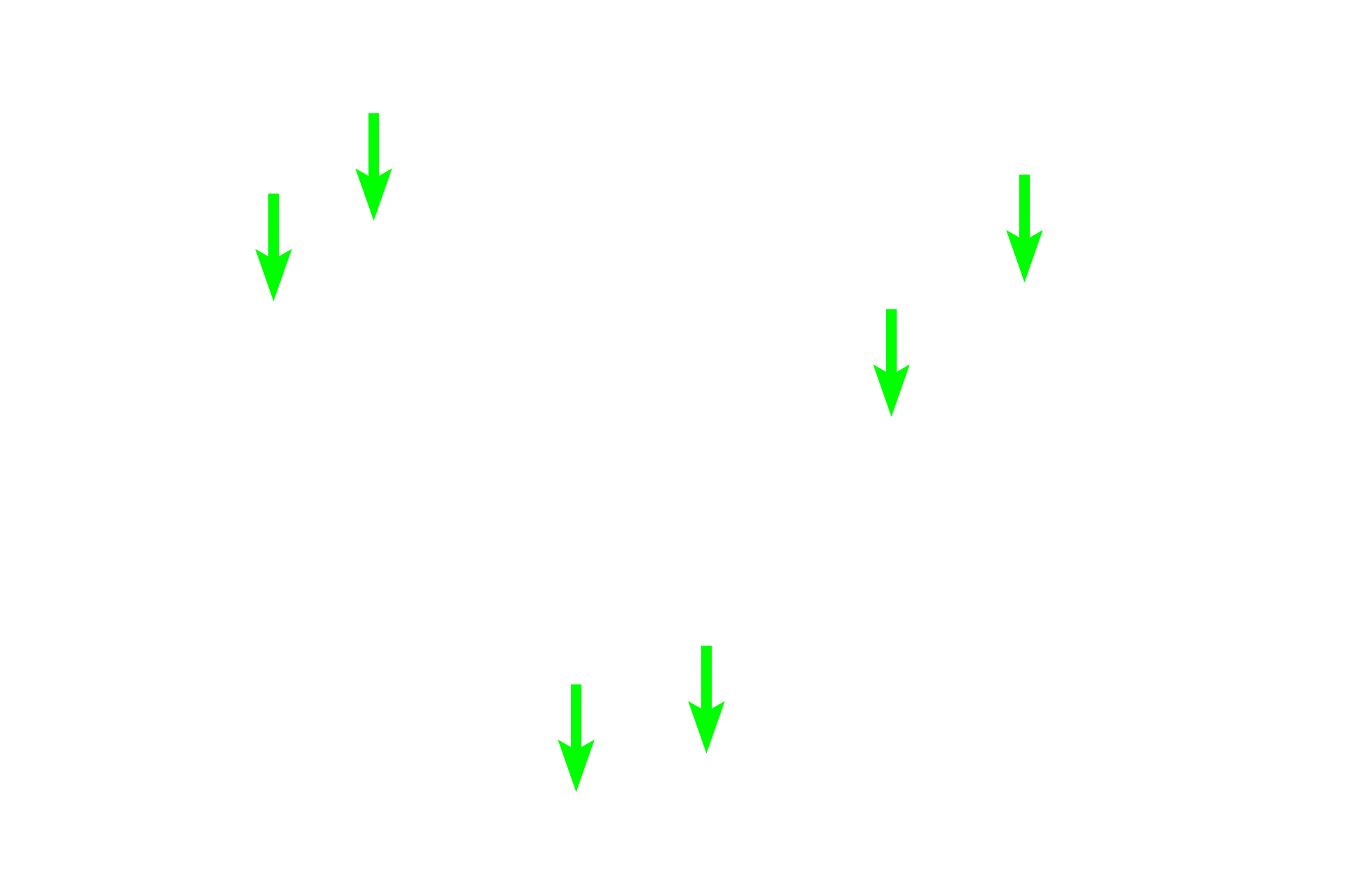
The body and tail of the epididymis are filled with the highly coiled duct of the epididymis. Numerous sectioned profiles of the single duct are seen in this image. The duct is lined by a very tall, regular, pseudostratified columnar epithelium with stereocilia, beneath which is layer of smooth muscle. As the duct progresses from the head to the tail region, its epithelium decreases in height and the smooth muscle layer increases in thickness. 40x

- Smooth muscle
The body and tail of the epididymis are filled with the highly coiled duct of the epididymis. Numerous sectioned profiles of the single duct are seen in this image. The duct is lined by a very tall, regular, pseudostratified columnar epithelium with stereocilia, beneath which is layer of smooth muscle. As the duct progresses from the head to the tail region, its epithelium decreases in height and the smooth muscle layer increases in thickness. 40x

Spermatoza
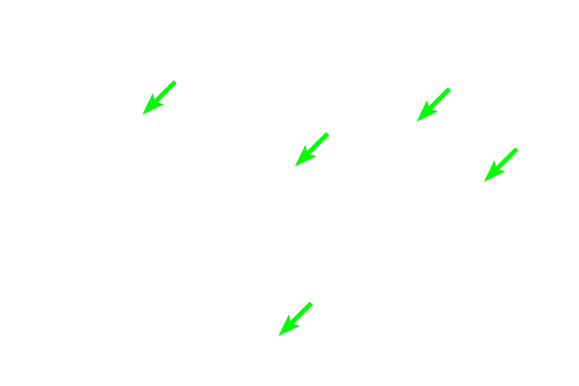
The body and tail of the epididymis are filled with the highly coiled duct of the epididymis. Numerous sectioned profiles of the single duct are seen in this image. The duct is lined by a very tall, regular, pseudostratified columnar epithelium with stereocilia, beneath which is layer of smooth muscle. As the duct progresses from the head to the tail region, its epithelium decreases in height and the smooth muscle layer increases in thickness. 40x