
Spleen: circulatory pathways
After leaving the white pulp, a central arteriole subdivides to form smaller arterioles and capillaries that eventually supply splenic sinuses in red pulp. The capillaries may open directly into the splenic sinuses (closed circulation) or indirectly by opening into splenic cords (open circulation). In the human, an open circulation is thought to predominate. 100x

Trabeculum
After leaving the white pulp, a central arteriole subdivides to form smaller arterioles and capillaries that eventually supply splenic sinuses in red pulp. The capillaries may open directly into the splenic sinuses (closed circulation) or indirectly by opening into splenic cords (open circulation). In the human an open circulation is thought to predominate. 100x

Periarterial lymphoid sheath (PALS)
After leaving the white pulp, a central arteriole subdivides to form smaller arterioles and capillaries that eventually supply splenic sinuses in red pulp. The capillaries may open directly into the splenic sinuses (closed circulation) or indirectly by opening into splenic cords (open circulation). In the human an open circulation is thought to predominate. 100x

Central arterioles
After leaving the white pulp, a central arteriole subdivides to form smaller arterioles and capillaries that eventually supply splenic sinuses in red pulp. The capillaries may open directly into the splenic sinuses (closed circulation) or indirectly by opening into splenic cords (open circulation). In the human an open circulation is thought to predominate. 100x

Lymphoid nodules
After leaving the white pulp, a central arteriole subdivides to form smaller arterioles and capillaries that eventually supply splenic sinuses in red pulp. The capillaries may open directly into the splenic sinuses (closed circulation) or indirectly by opening into splenic cords (open circulation). In the human an open circulation is thought to predominate. 100x

Splenic sinuses
After leaving the white pulp, a central arteriole subdivides to form smaller arterioles and capillaries that eventually supply splenic sinuses in red pulp. The capillaries may open directly into the splenic sinuses (closed circulation) or indirectly by opening into splenic cords (open circulation). In the human an open circulation is thought to predominate. 100x

Splenic cords
After leaving the white pulp, a central arteriole subdivides to form smaller arterioles and capillaries that eventually supply splenic sinuses in red pulp. The capillaries may open directly into the splenic sinuses (closed circulation) or indirectly by opening into splenic cords (open circulation). In the human an open circulation is thought to predominate. 100x
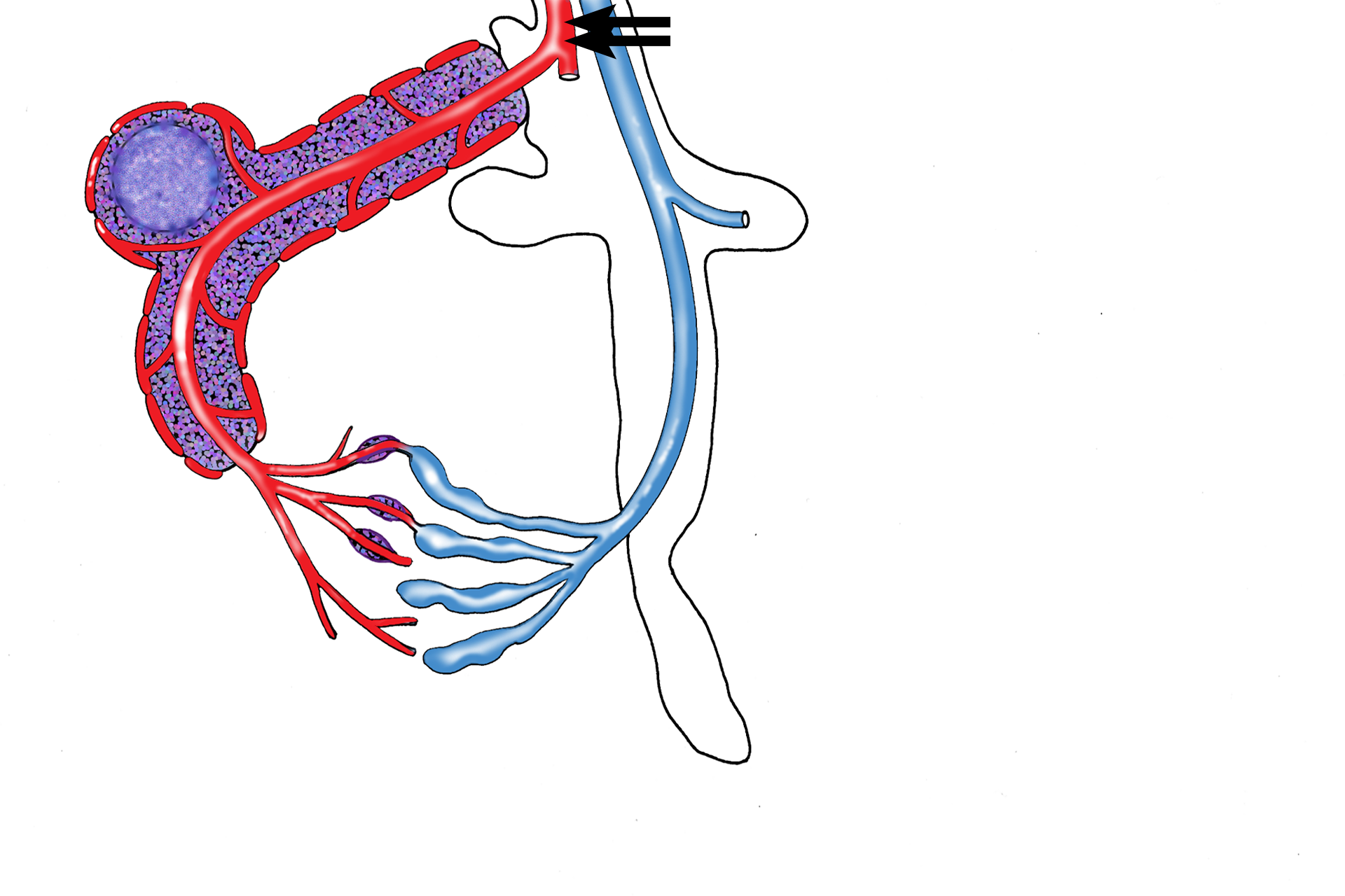
Trabecular artery >
The splenic artery enters the spleen and subdivides into branches that enter trabeculae, called trabecular arteries.
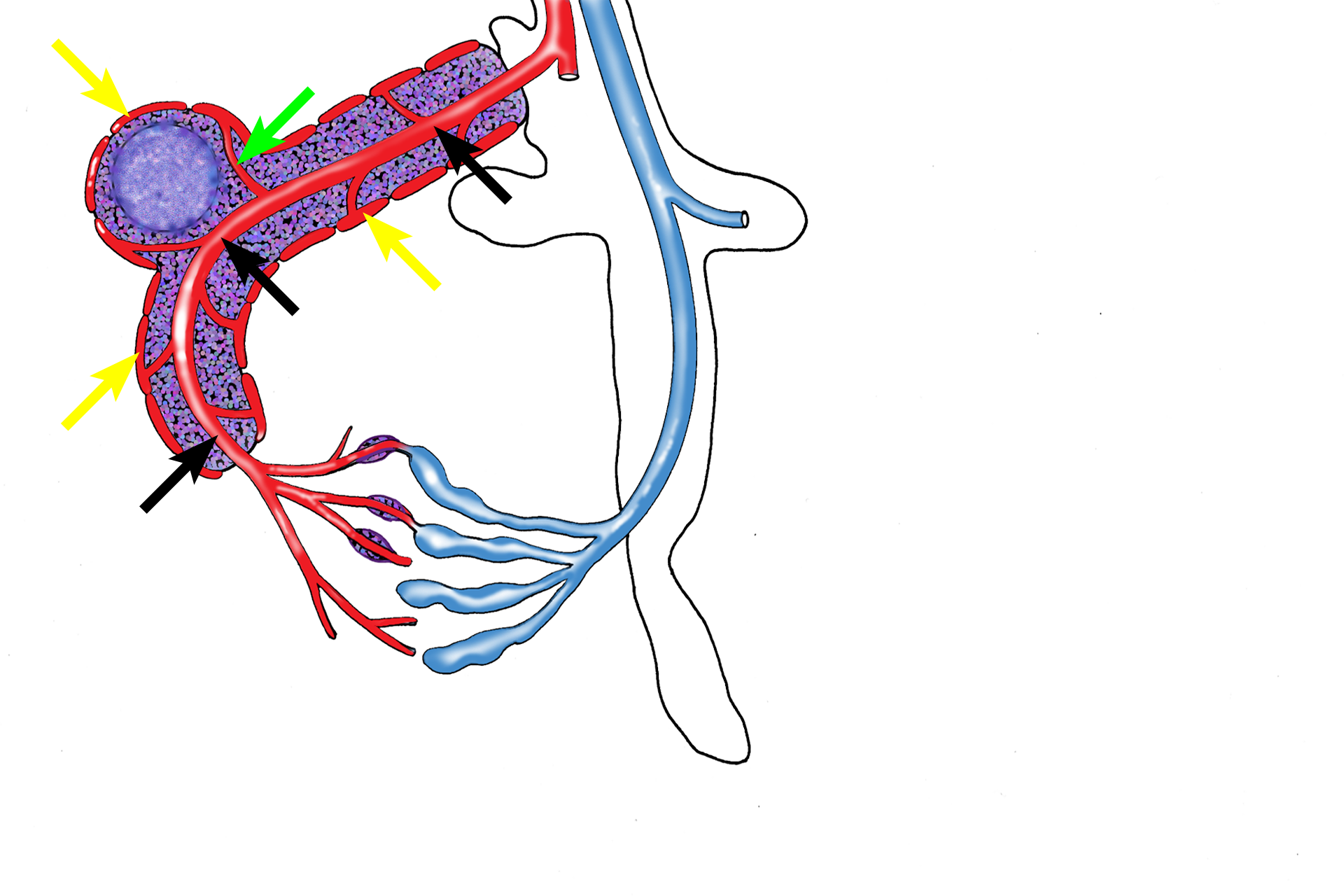
White pulp vasculature >
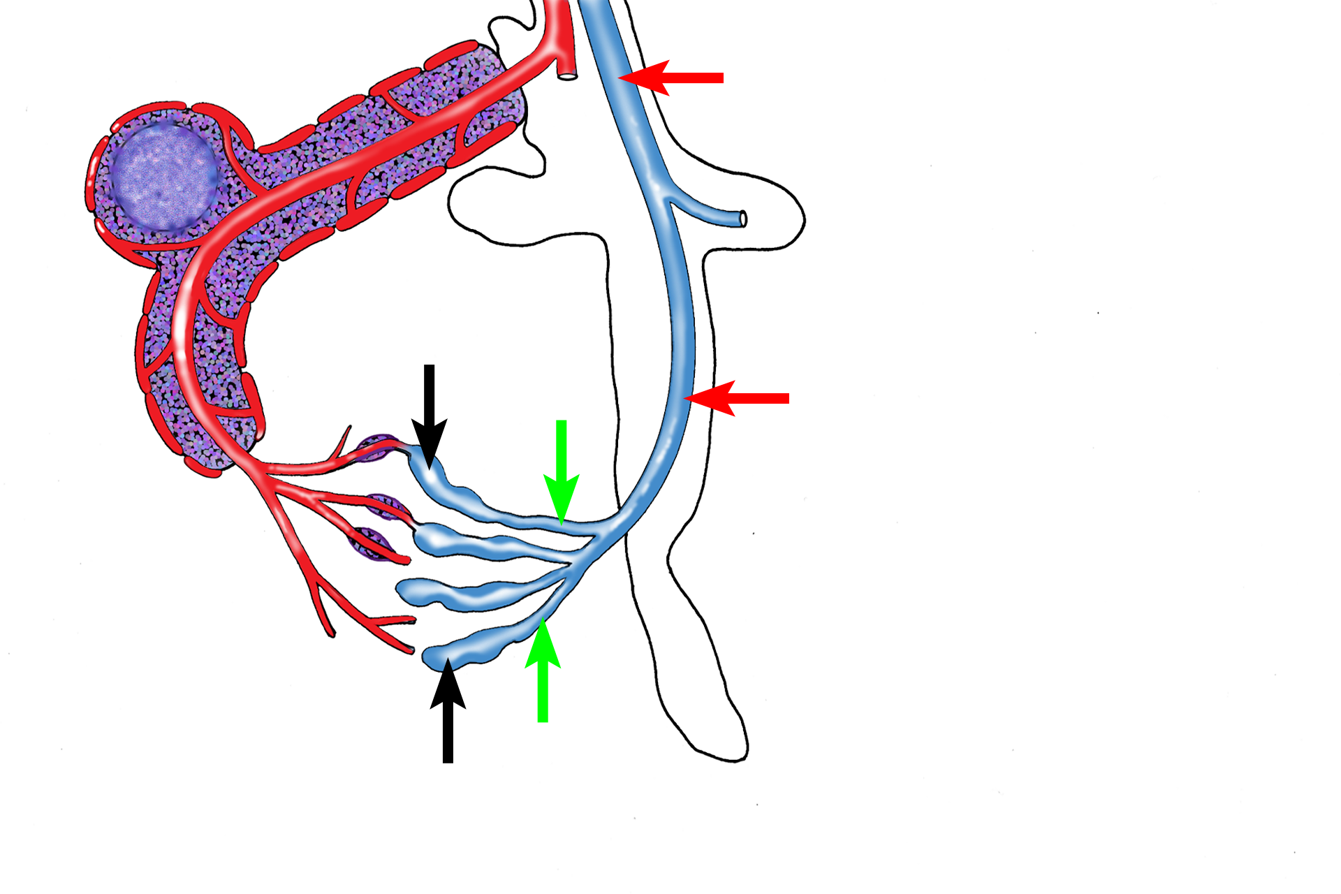
When arterioles leave the trabeculae to enter the white pulp, they are called central arterioles (black arrows) and are surrounded by PALS; branches of these vessels supply the PALS and lymphoid nodules (green arrow) and marginal sinuses (yellow arrows).
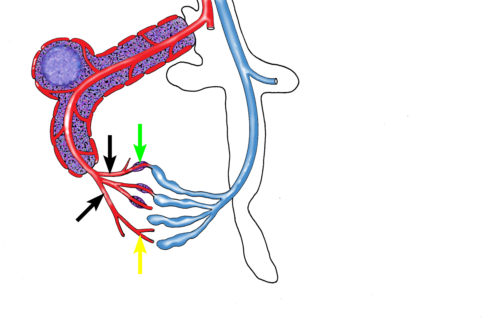
Red pulp vasculature >
When the central arteriole leaves the white pulp to enter the red pulp, it divides into a series of smaller arterioles, called penicillar arteries (black arrows), which continue as sheathed (green arrows) and unsheathed (yellow arrows) capillaries.
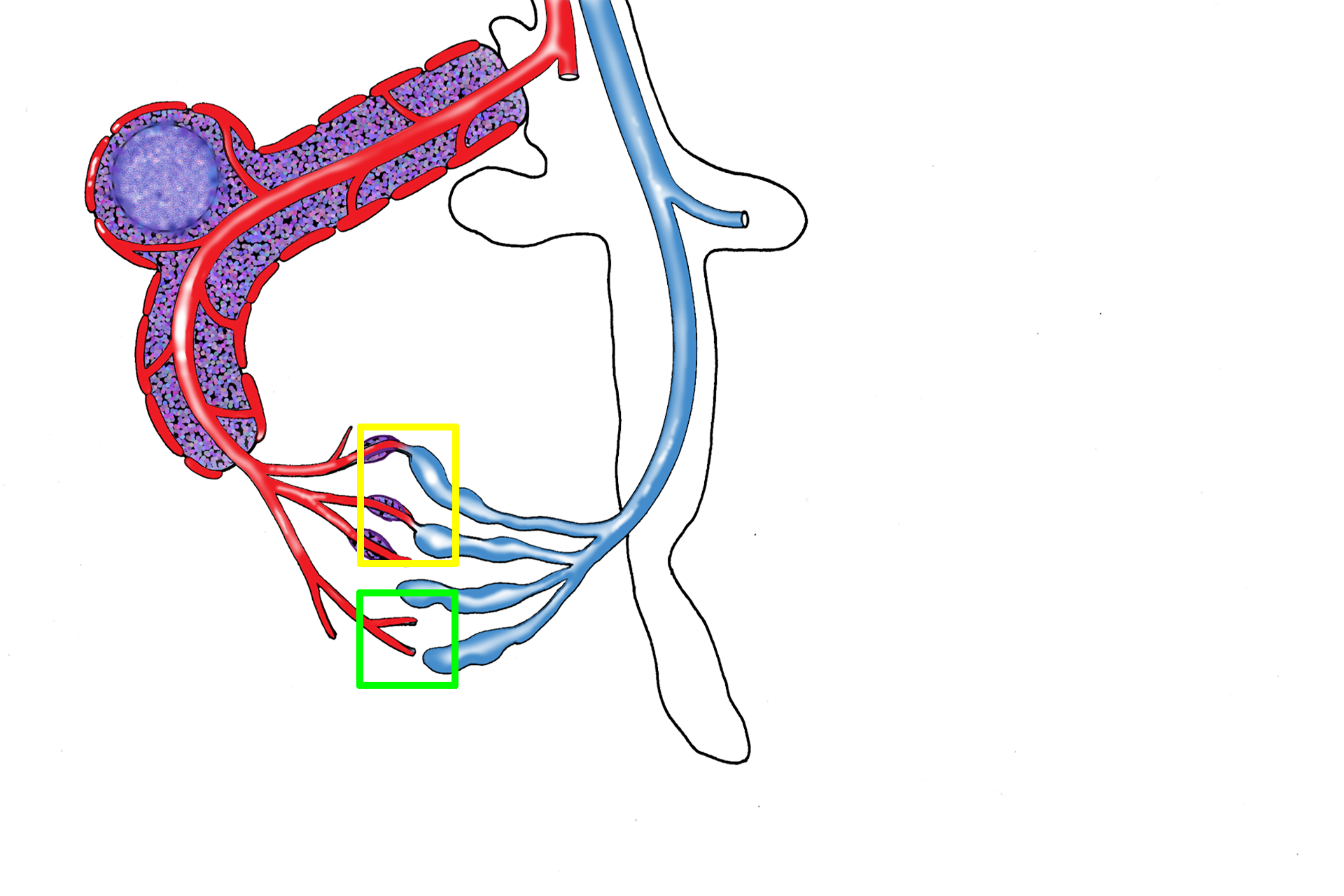
Open/closed circulations >
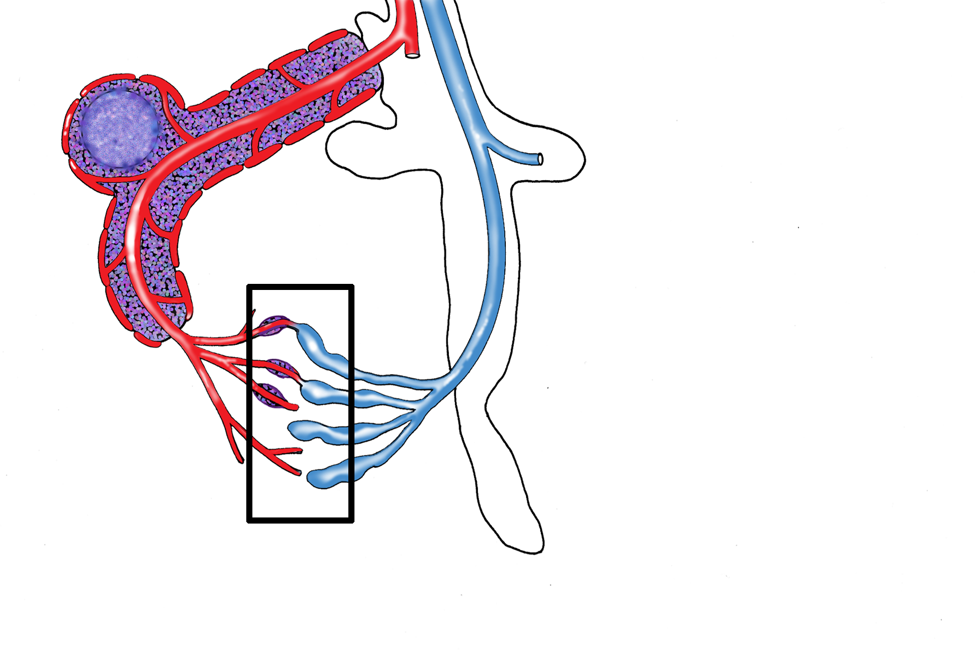
Sheathed and unsheathed capillaries either connect directly with the splenic sinuses (closed circulation, yellow rectangle) or open directly into the reticular connective tissue of the splenic cord, allowing blood to be exposed to cells of the splenic cord (open circulation, green square). This open pathway allows blood to percolate indirectly into the splenic sinuses and is thought to predominate in humans.
Venous drainage >
Splenic sinuses (black arrows) anastomose to form splenic veins (green arrows) that enter the trabeculae as trabecular veins (red arrows). Trabecular veins anastomose to form the splenic vein, which exits from the spleen at the hilum.
Area shown in next image >
The next image illustrates the open and closed circulations within the splenic red pulp.