
Stroma and parenchyma
These images show parenchymal and stromal elements at higher magnification in the stomach and small intestine. 400x (stomach, H&E, left), 100x (small intestine, trichrome, right)
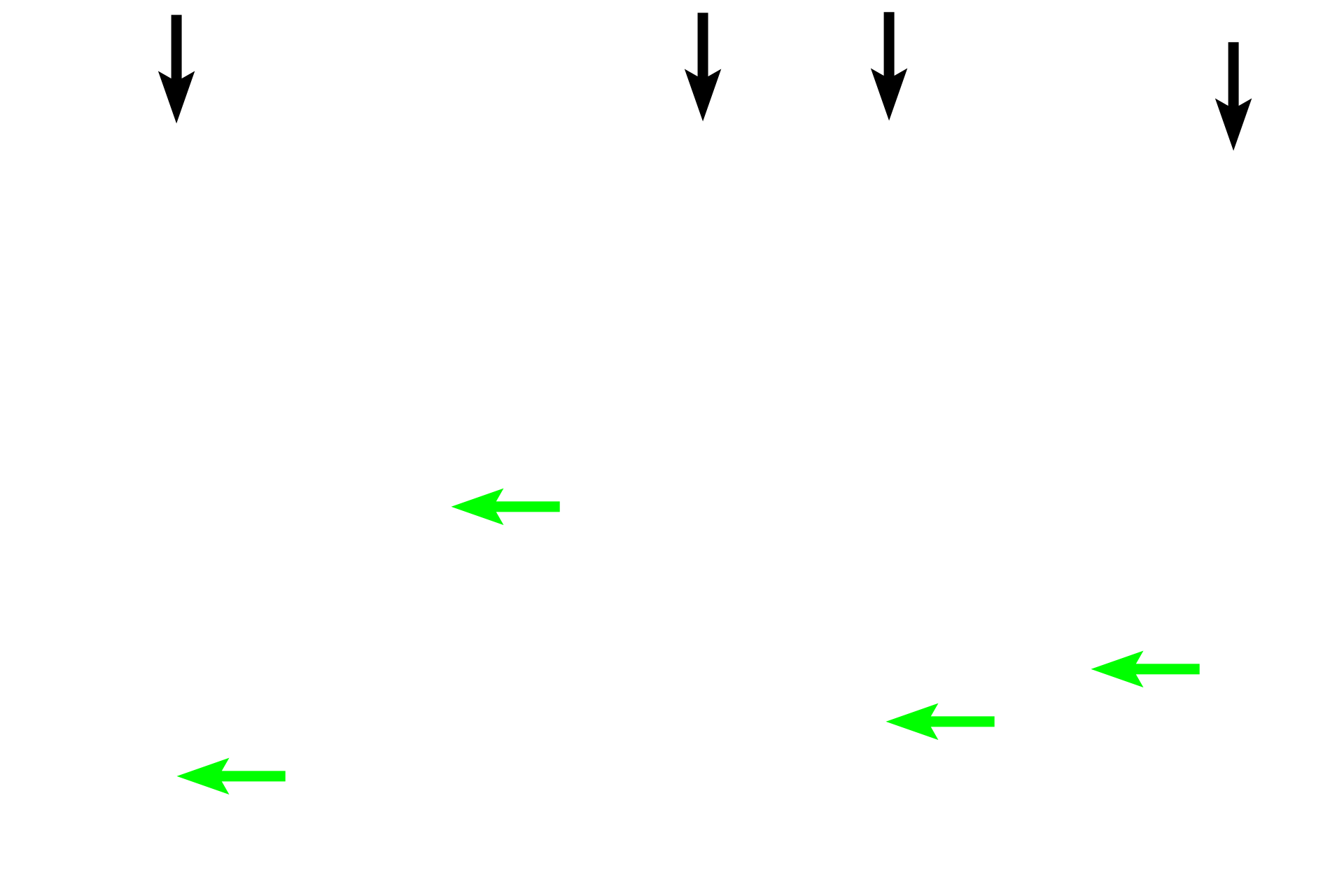
Parenchyma >
In the mucosa of these two organs, the surface epithelium (black arrows) and glands (green arrows) form the parenchyma.
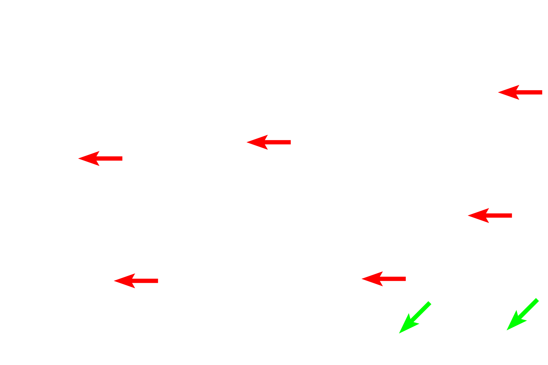
Stroma >
The stroma of an organ is composed of connective tissues both loose (red arrows) and dense (green arrows).
- Loose connective tissue >
A loose connective tissue stroma is present beneath the surface epithelium and surrounding the glands. The gelatinous ground substance of this stroma provides good padding for the epithelium, supports the glands and allows for diffusion and cell migration. Delicate collagen fibers are the predominant fiber type in this stroma.

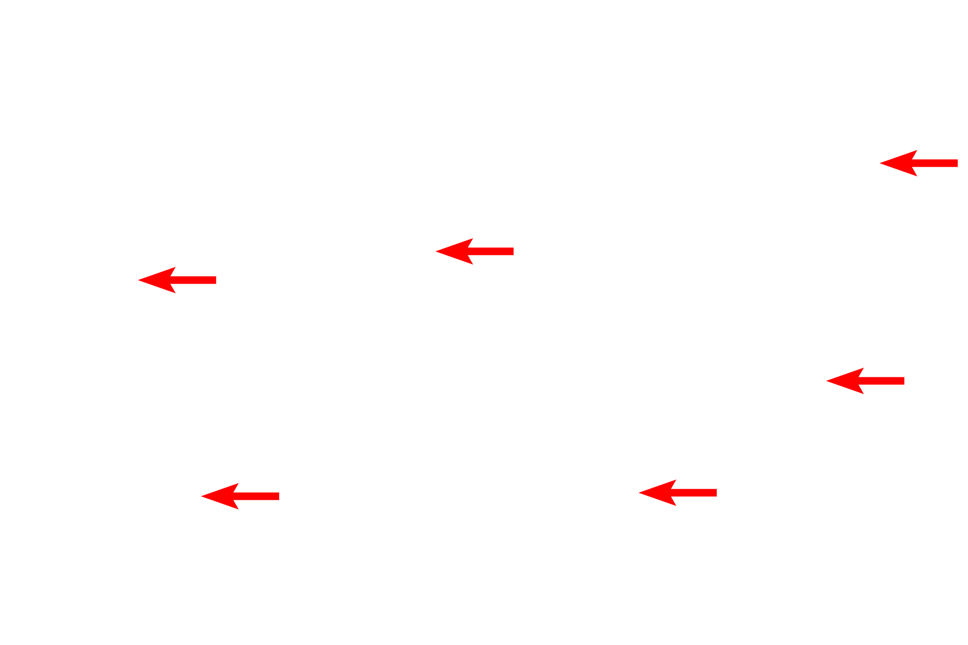
- Dense connective tissue >
The stroma also provides more robust structural support for organs wall. In these areas, thick collagen fibers of the dense connective tissue predominate. This section is stained by the trichrome method which demonstrates the stroma of an organ by labeling the connective tissue bluish green.
Lumen
These images show parenchymal and stromal elements at higher magnification in the stomach and small intestine. 400x (stomach, H&E, left), 100x (small intestine, trichrome, right)