
Sensory retina
This image compares two images of the retina at a similar magnification. The image in the left panel was generated by Dr. Johannes A. G. Rhodin, “An Atlas of Histology” (Oxford Press, 1974) and maintained in the University of Michigan collection. 400x, 400x

Retina
This image compares the appearance of the retina seen by light microscopy (right) and electron microscopy (left) at a similar magnification. 400x

- Pigment epithelium
This image compares the appearance of the retina seen by light microscopy (right) and electron microscopy (left) at a similar magnification. 400x

- Photoreceptor layer
This image compares the appearance of the retina seen by light microscopy (right) and electron microscopy (left) at a similar magnification. 400x
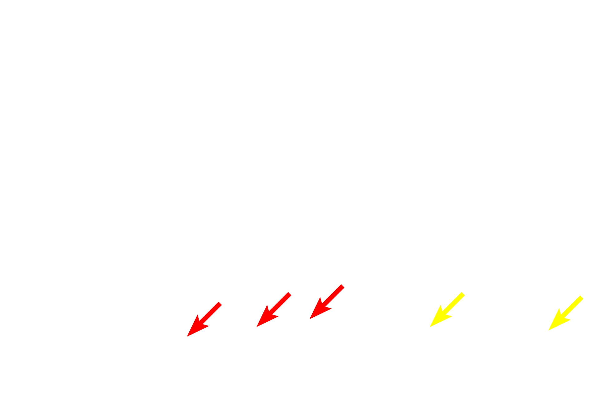
-- Outer segments of rods and cones
This image compares the appearance of the retina seen by light microscopy (right) and electron microscopy (left) at a similar magnification. 400x
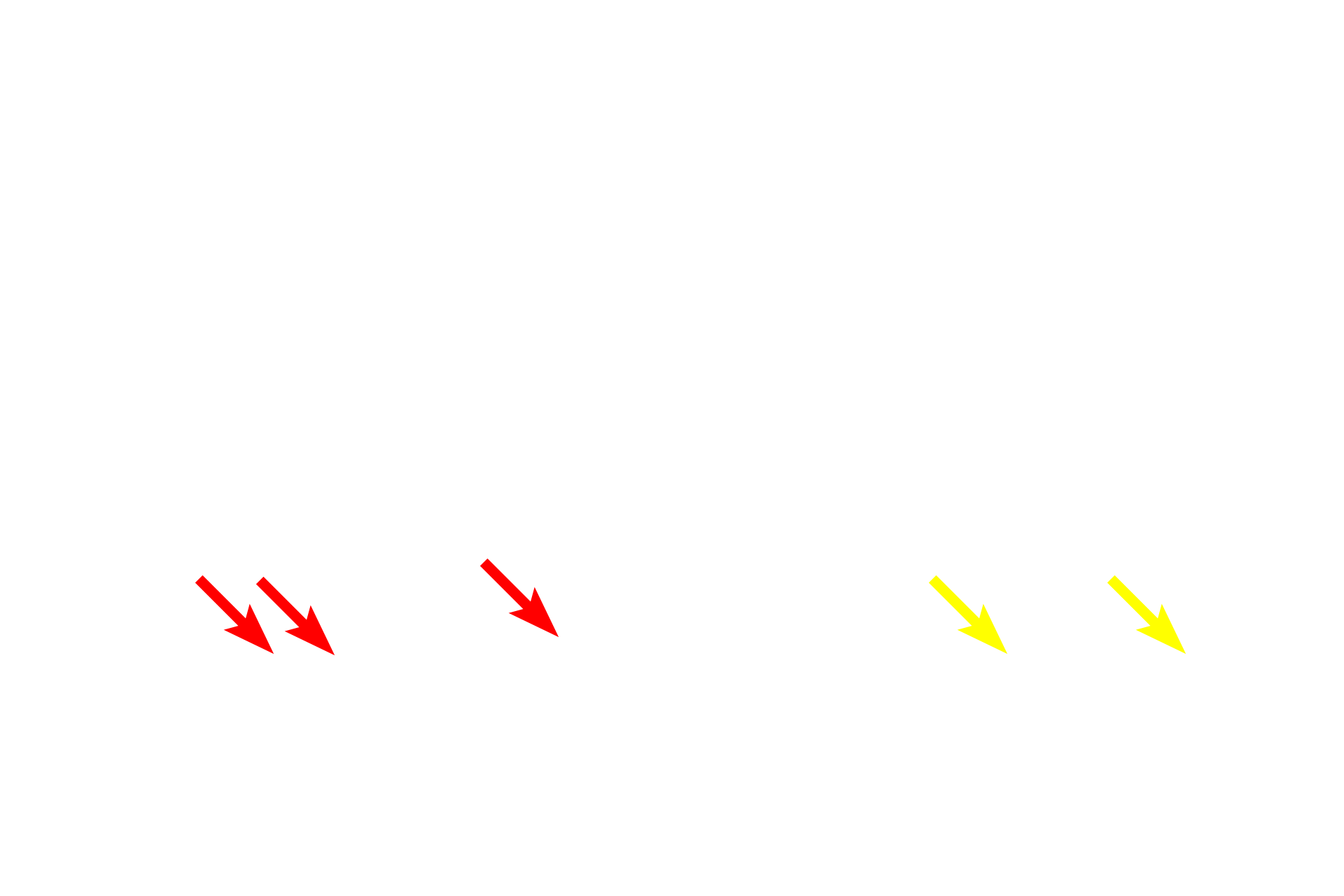
-- Inner segments of rods and cones
This image compares the appearance of the retina seen by light microscopy (right) and electron microscopy (left) at a similar magnification. 400x

-- Outer limiting membrane
This image compares the appearance of the retina seen by light microscopy (right) and electron microscopy (left) at a similar magnification. 400x

-- Outer nuclear layer
This image compares the appearance of the retina seen by light microscopy (right) and electron microscopy (left) at a similar magnification. 400x
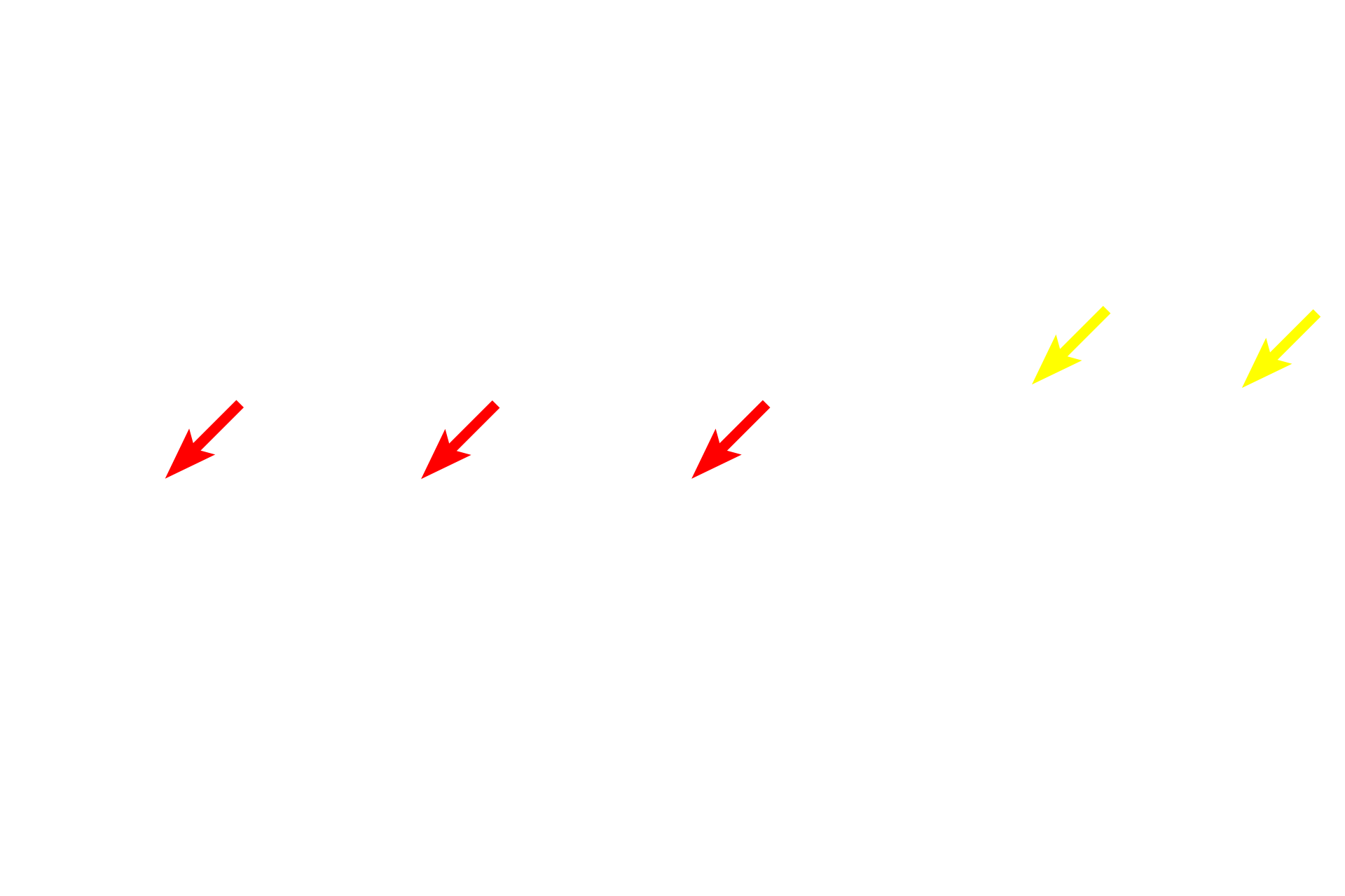
- Outer plexiform layer
This image compares the appearance of the retina seen by light microscopy (right) and electron microscopy (left) at a similar magnification. 400x

- Inner nuclear layer
This image compares the appearance of the retina seen by light microscopy (right) and electron microscopy (left) at a similar magnification. 400x

- Inner plexiform layer
This image compares the appearance of the retina seen by light microscopy (right) and electron microscopy (left) at a similar magnification. 400x

- Ganglion cell layer
This image compares the appearance of the retina seen by light microscopy (right) and electron microscopy (left) at a similar magnification. 400x
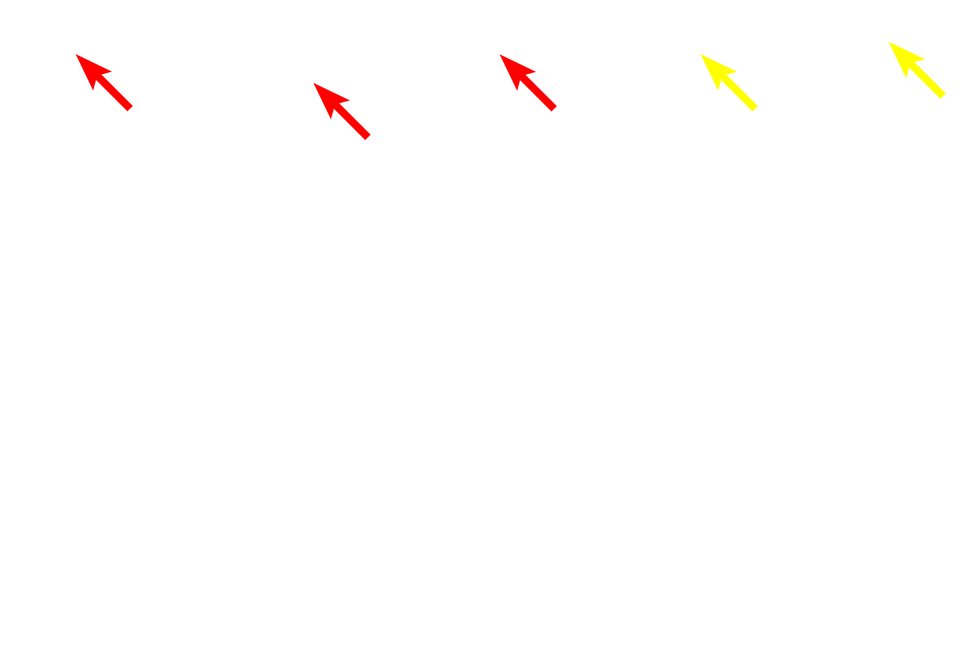
- Nerve fiber layer
This image compares the appearance of the retina seen by light microscopy (right) and electron microscopy (left) at a similar magnification. 400x

- Inner limiting membrane
This image compares the appearance of the retina seen by light microscopy (right) and electron microscopy (left) at a similar magnification. 400x

Choroid
This image compares the appearance of the retina seen by light microscopy (right) and electron microscopy (left) at a similar magnification. 400x

Choriocapillaris
This image compares the appearance of the retina seen by light microscopy (right) and electron microscopy (left) at a similar magnification. 400x

Bruch’s membrane
This image compares the appearance of the retina seen by light microscopy (right) and electron microscopy (left) at a similar magnification. 400x
Image source >
This electron micrograph, originally published by J.A.G. Rhodin, some from the University of Michigan collection.