
Semicircular canals
Sections through the inner ear demonstrate two of its three major subdivisions: the osseous vestibule with its membranous utricle and saccule and their receptors, the maculae; three osseous semicircular canals with their membranous semicircular ducts and their receptors, the crista ampullares.

Plane of section - #1
Sections through the inner ear demonstrate two of its three major subdivisions: the osseous vestibule with its membranous utricle and saccule and their receptors, the maculae; three osseous semicircular canals with their membranous semicircular ducts and their receptors, the crista ampullares.

Plane of section - #2
Sections through the inner ear demonstrate two of its three major subdivisions: the osseous vestibule with its membranous utricle and saccule and their receptors, the maculae; three osseous semicircular canals with their membranous semicircular ducts and their receptors, the crista ampullares.
Plane of section - #3
Sections through the inner ear demonstrate two of its three major subdivisions: the osseous vestibule with its membranous utricle and saccule and their receptors, the maculae; three osseous semicircular canals with their membranous semicircular ducts and their receptors, the crista ampullares.
Temporal bone, petrous >
The inner ear is nested in and protected by the petrous portion of the temporal bone.

Vestibule >
The vestibule, a portion of the osseous labyrinth, houses the utricle and saccule, portions of the membranous labyrinth. Receptors in the utricle and saccule, the maculae, respond to linear acceleration and gravitational forces.
- Utricle
The vestibule, a portion of the osseous labyrinth, houses the utricle and saccule, portions of the membranous labyrinth. Receptors in the utricle and saccule, the maculae, respond to linear acceleration and gravitational forces.
- Saccule
The vestibule, a portion of the osseous labyrinth, houses the utricle and saccule, portions of the membranous labyrinth. Receptors in the utricle and saccule, the maculae, respond to linear acceleration and gravitational forces.
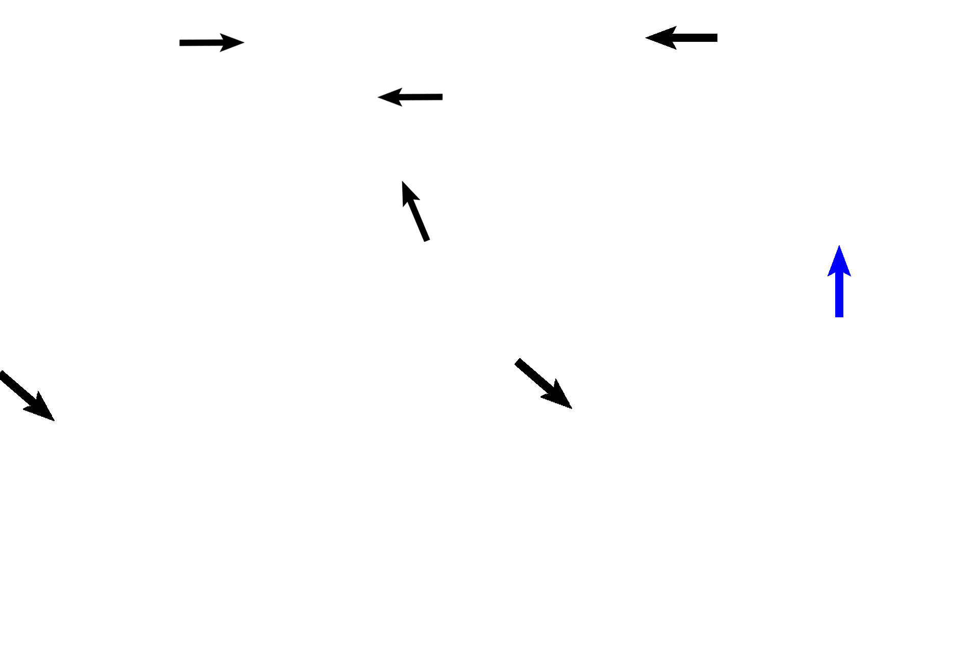
Semicircular canals >
Portions of three semicircular canals (black arrows, osseous labyrinth), are shown at or near their connections with the vestibule. The fourth canal (blue arrow) is shown extending some distance from the vestibule.
- Semicircular ducts >
A semicircular duct, part of the membranous labyrinth, is suspended in each semicircular canal. Each duct possesses a crista ampullaris (only one is visible), the receptor that responds to angular acceleration.

-- Crista ampullaris
A semicircular duct, part of the membranous labyrinth, is suspended in each semicircular canal. Each duct possesses a crista ampullaris (only one is visible), the receptor that responds to angular acceleration.
Connective tissue reticulum >
The connective tissue reticulum secures the membranous labyrinth in the osseous labyrinth.

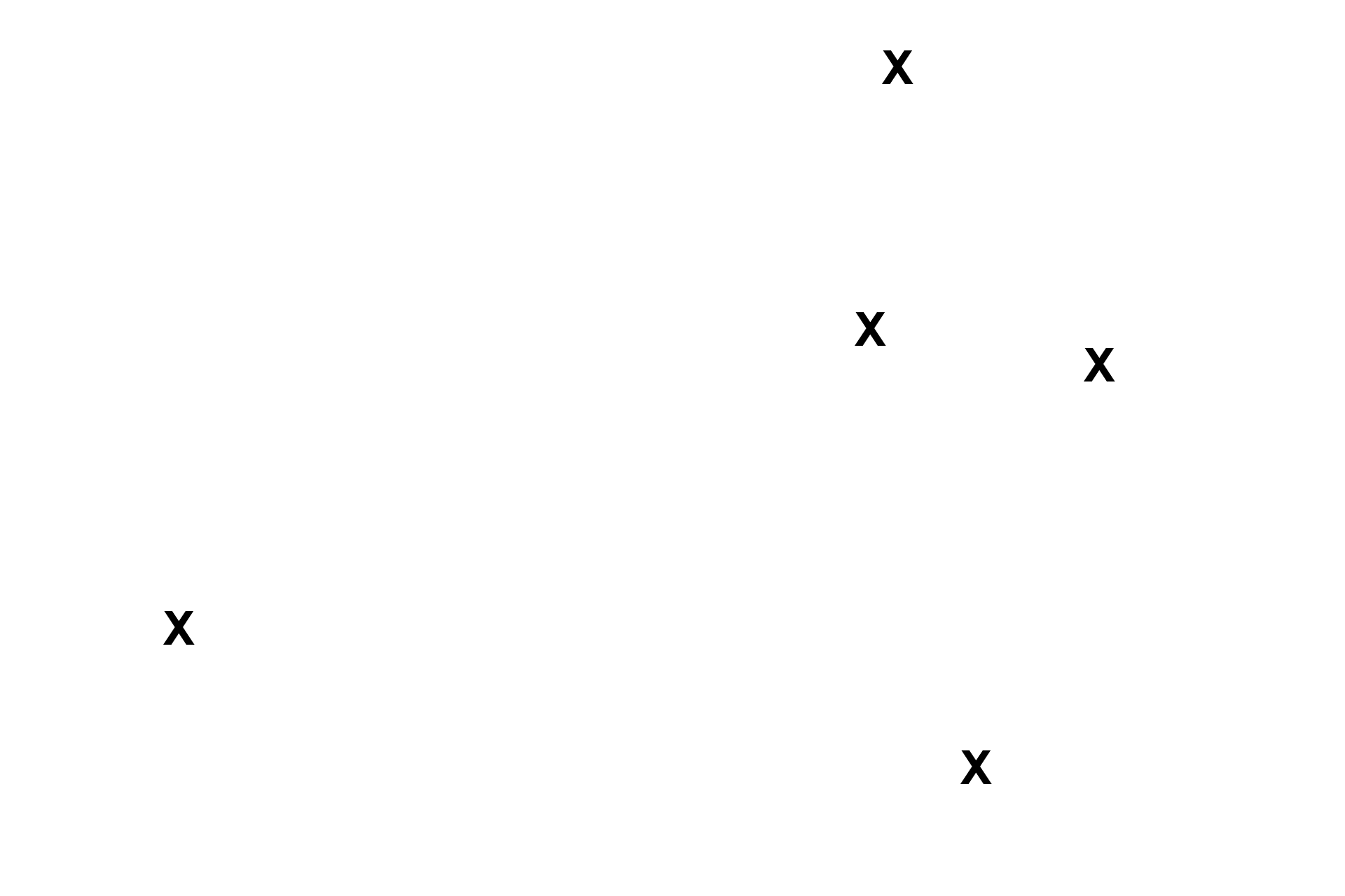
Middle ear
A portion of the middle ear (x) shows the foot plate of the stapes (arrow) inserting into the oval window separating the middle ear from the vestibule.
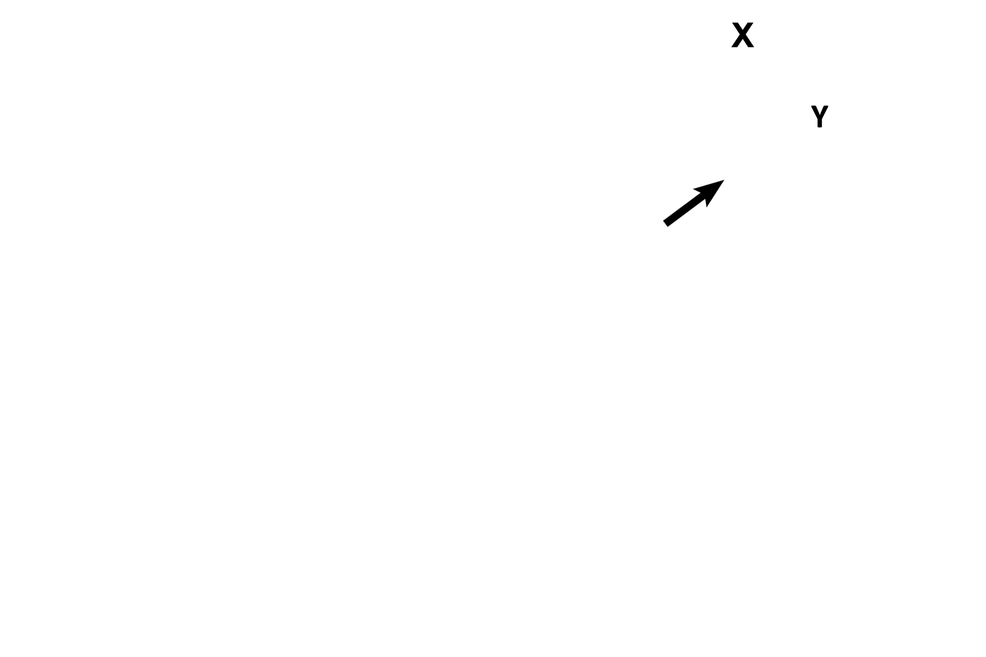
Cranial cavity and brain
The cranial cavity (X) and brain (Y) are separated from the ear only by a thin layer of temporal bone (arrow).