![This is blank.](https://digitalhistology.org/wp-content/themes/histology-unified/imgs/trans.png)
Large intestine
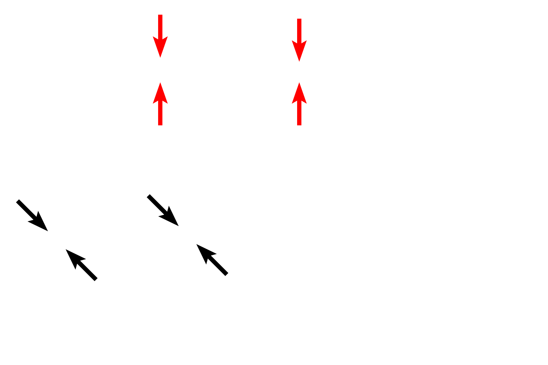
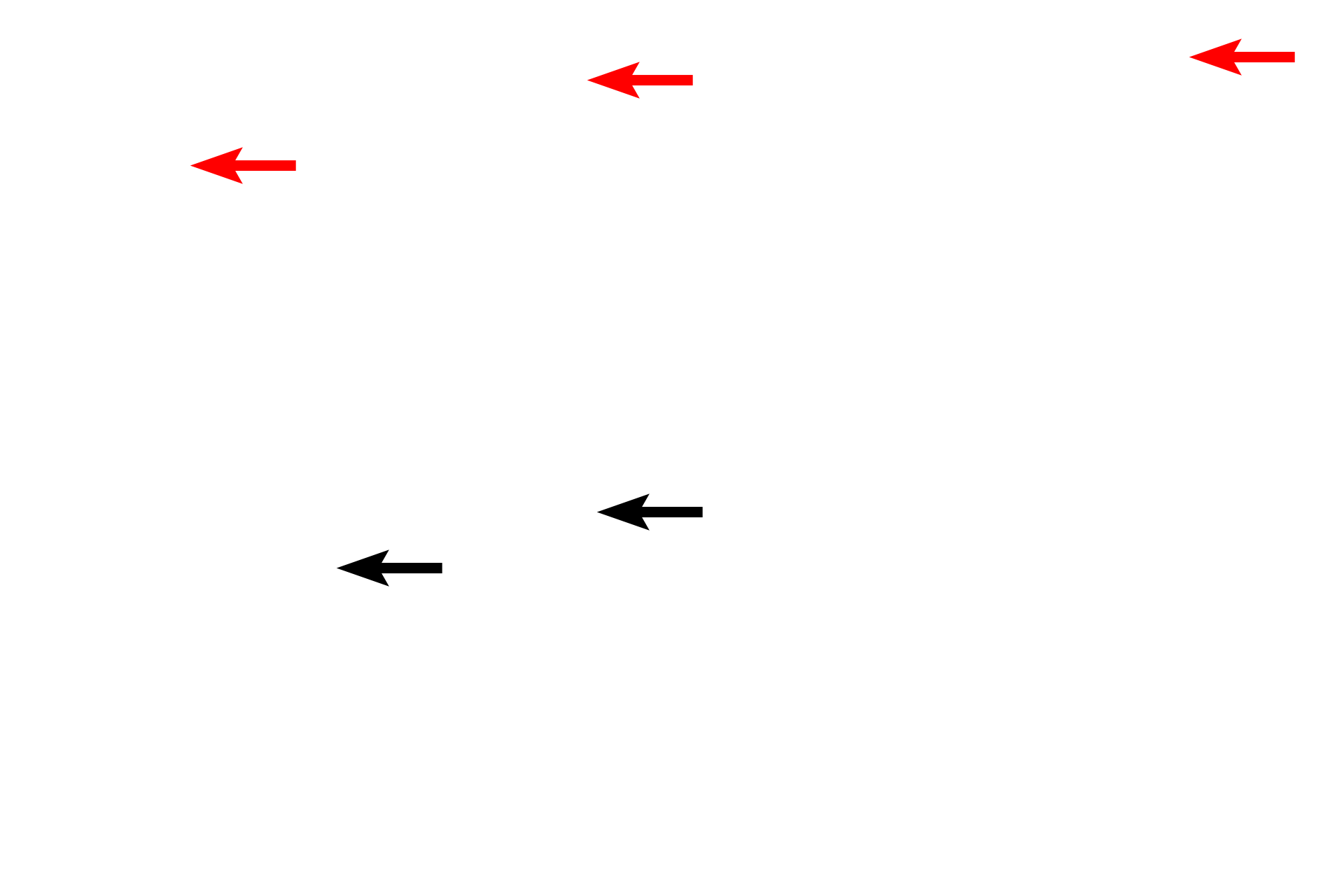
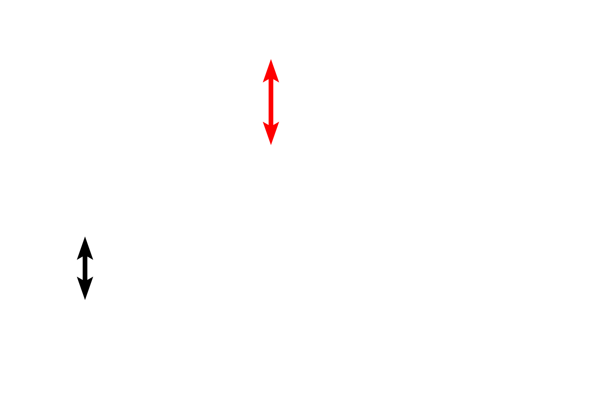
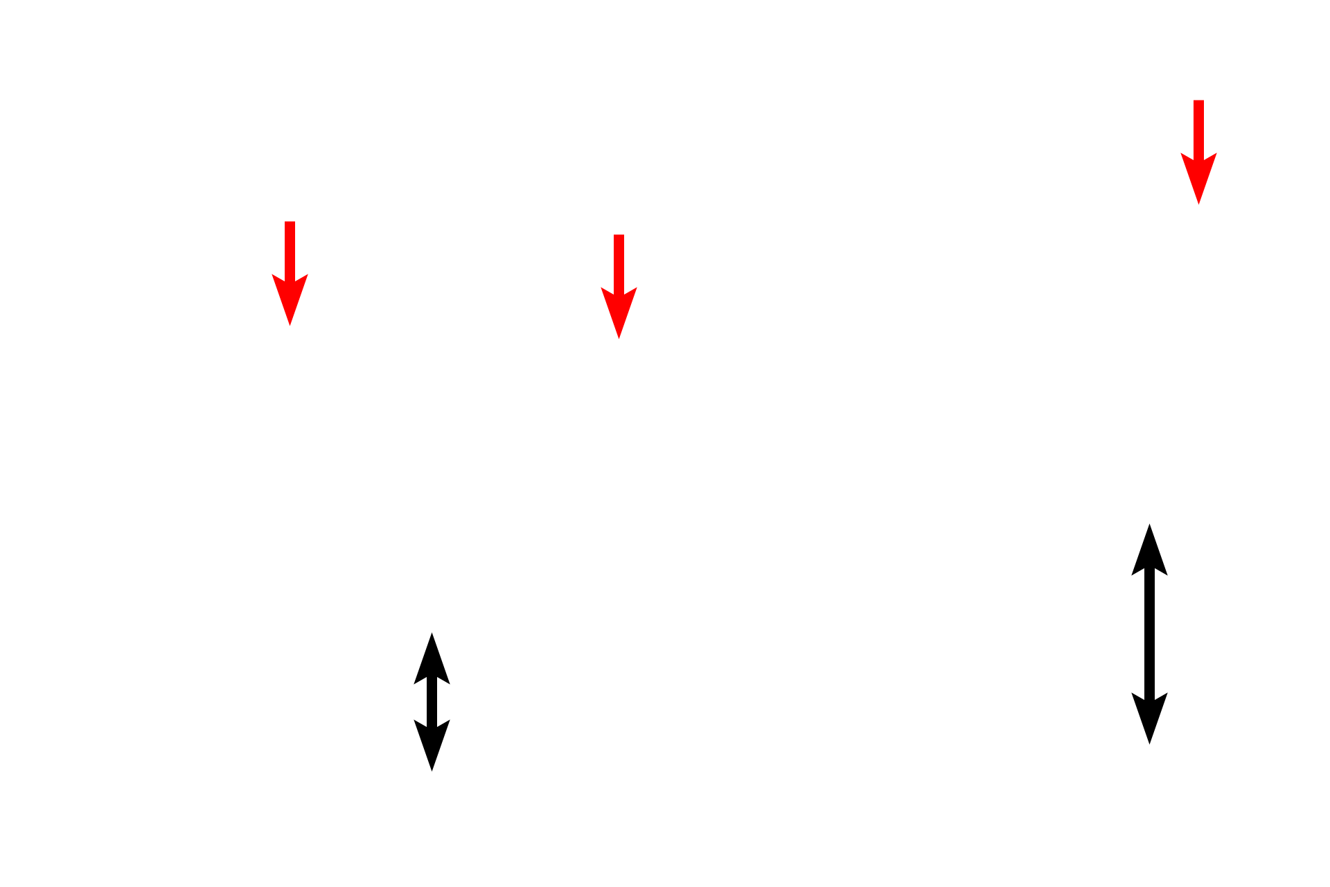
The large intestine is composed of the four basic tunics found throughout the tubular digestive system. It is differentiated from small intestine by it overall flat surface, lack of villi, straight intestinal glands and taeniae coli. The large intestine is the site of water absorption and fecal formation. The large intestine was cut in cross section in the top left images and longitudinally through a taenia coli in the bottom images. 10x, 10x, 100x, 100x
![Mucosa <p>The large intestine is composed of the four basic tunics found throughout the tubular digestive system. It is differentiated from small intestine by it overall flat surface, lack of villi, straight intestinal glands and taeniae coli. The large intestine is the site of water absorption and fecal formation. The large intestine was cut in cross section in the top left images and longitudinally through a taenia coli in the bottom images. 10x, 10x, 100x, 100x</p>](https://digitalhistology.org/wp-content/uploads/2024/04/Or.Dig_.Tub_.LgI-2A-1-ping.png)
Mucosa
The large intestine is composed of the four basic tunics found throughout the tubular digestive system. It is differentiated from small intestine by it overall flat surface, lack of villi, straight intestinal glands and taeniae coli. The large intestine is the site of water absorption and fecal formation. The large intestine was cut in cross section in the top left images and longitudinally through a taenia coli in the bottom images. 10x, 10x, 100x, 100x
![- Intestinal glands <p>The large intestine is composed of the four basic tunics found throughout the tubular digestive system. It is differentiated from small intestine by it overall flat surface, lack of villi, straight intestinal glands and taeniae coli. The large intestine is the site of water absorption and fecal formation. The large intestine was cut in cross section in the top left images and longitudinally through a taenia coli in the bottom images. 10x, 10x, 100x, 100x</p>](https://digitalhistology.org/wp-content/uploads/2024/04/Or.Dig_.Tub_.LgI-2A-7-ping.png)
- Intestinal glands
The large intestine is composed of the four basic tunics found throughout the tubular digestive system. It is differentiated from small intestine by it overall flat surface, lack of villi, straight intestinal glands and taeniae coli. The large intestine is the site of water absorption and fecal formation. The large intestine was cut in cross section in the top left images and longitudinally through a taenia coli in the bottom images. 10x, 10x, 100x, 100x
![Submucosa <p>The large intestine is composed of the four basic tunics found throughout the tubular digestive system. It is differentiated from small intestine by it overall flat surface, lack of villi, straight intestinal glands and taeniae coli. The large intestine is the site of water absorption and fecal formation. The large intestine was cut in cross section in the top left images and longitudinally through a taenia coli in the bottom images. 10x, 10x, 100x, 100x</p>](https://digitalhistology.org/wp-content/uploads/2024/04/Or.Dig_.Tub_.LgI-2A-2-ping.png)
Submucosa
The large intestine is composed of the four basic tunics found throughout the tubular digestive system. It is differentiated from small intestine by it overall flat surface, lack of villi, straight intestinal glands and taeniae coli. The large intestine is the site of water absorption and fecal formation. The large intestine was cut in cross section in the top left images and longitudinally through a taenia coli in the bottom images. 10x, 10x, 100x, 100x
![Inner muscularis externa <p>The large intestine is composed of the four basic tunics found throughout the tubular digestive system. It is differentiated from small intestine by it overall flat surface, lack of villi, straight intestinal glands and taeniae coli. The large intestine is the site of water absorption and fecal formation. The large intestine was cut in cross section in the top left images and longitudinally through a taenia coli in the bottom images. 10x, 10x, 100x, 100x</p>](https://digitalhistology.org/wp-content/uploads/2024/04/Or.Dig_.Tub_.LgI-2A-3-ping.png)
Inner muscularis externa
The large intestine is composed of the four basic tunics found throughout the tubular digestive system. It is differentiated from small intestine by it overall flat surface, lack of villi, straight intestinal glands and taeniae coli. The large intestine is the site of water absorption and fecal formation. The large intestine was cut in cross section in the top left images and longitudinally through a taenia coli in the bottom images. 10x, 10x, 100x, 100x
![Outer muscularis externa <p>The large intestine is composed of the four basic tunics found throughout the tubular digestive system. It is differentiated from small intestine by it overall flat surface, lack of villi, straight intestinal glands and taeniae coli. The large intestine is the site of water absorption and fecal formation. The large intestine was cut in cross section in the top left images and longitudinally through a taenia coli in the bottom images. 10x, 10x, 100x, 100x</p>](https://digitalhistology.org/wp-content/uploads/2024/04/Or.Dig_.Tub_.LgI-2A-4-ping.png)
Outer muscularis externa
The large intestine is composed of the four basic tunics found throughout the tubular digestive system. It is differentiated from small intestine by it overall flat surface, lack of villi, straight intestinal glands and taeniae coli. The large intestine is the site of water absorption and fecal formation. The large intestine was cut in cross section in the top left images and longitudinally through a taenia coli in the bottom images. 10x, 10x, 100x, 100x
![- Taeniae coli > <p>The majority of the outer longitudinal portion of muscularis externa is segregated into three separate bands called taeniae coli, seen cut in cross section in the upper image. In the bottom image, the muscularis externa has been cut longitudinally through a taenia coli, demonstrating its thickness.</p>](https://digitalhistology.org/wp-content/uploads/2024/04/Or.Dig_.Tub_.LgI-2A-5-ping.png)
- Taeniae coli >
The majority of the outer longitudinal portion of muscularis externa is segregated into three separate bands called taeniae coli, seen cut in cross section in the upper image. In the bottom image, the muscularis externa has been cut longitudinally through a taenia coli, demonstrating its thickness.
![Serosa > <p>The outermost layer of the large intestine is either an adventitia or a serosa, depending on whether a particular segment of this organ is located intra- or retroperitoneally.</p>](https://digitalhistology.org/wp-content/uploads/2024/04/Or.Dig_.Tub_.LgI-2A-6-ping.png)
Serosa >
The outermost layer of the large intestine is either an adventitia or a serosa, depending on whether a particular segment of this organ is located intra- or retroperitoneally.