
Recto-anal junction
The anus is seen in the right half of this image. It is lined by a stratified squamous, keratinized epithelium and possesses apocrine sweat glands, eccrine sweat glands, sebaceous glands and hair follicles. Apocrine sweat glands are characterized by their large size, tubular structure and wide lumens. Also visible are the internal and external anal sphincters. 40x
Rectum
The anus is seen in the right half of this image. It is lined by a stratified squamous, keratinized epithelium and possesses apocrine sweat glands, eccrine sweat glands, sebaceous glands and hair follicles. Apocrine sweat glands are characterized by their large size, tubular structure and wide lumens. Also visible are the internal and external anal sphincters. 40x

Recto-anal junction
The anus is seen in the right half of this image. It is lined by a stratified squamous, keratinized epithelium and possesses apocrine sweat glands, eccrine sweat glands, sebaceous glands and hair follicles. Apocrine sweat glands are characterized by their large size, tubular structure and wide lumens. Also visible are the internal and external anal sphincters. 40x

Anal canal
The anus is seen in the right half of this image. It is lined by a stratified squamous, keratinized epithelium and possesses apocrine sweat glands, eccrine sweat glands, sebaceous glands and hair follicles. Apocrine sweat glands are characterized by their large size, tubular structure and wide lumens. Also visible are the internal and external anal sphincters. 40x
Anus
The anus is seen in the right half of this image. It is lined by a stratified squamous, keratinized epithelium and possesses apocrine sweat glands, eccrine sweat glands, sebaceous glands and hair follicles. Apocrine sweat glands are characterized by their large size, tubular structure and wide lumens. Also visible are the internal and external anal sphincters. 40x
Apocrine sweat glands
The anus is seen in the right half of this image. It is lined by a stratified squamous, keratinized epithelium and possesses apocrine sweat glands, eccrine sweat glands, sebaceous glands and hair follicles. Apocrine sweat glands are characterized by their large size, tubular structure and wide lumens. Also visible are the internal and external anal sphincters. 40x

Hair follicles
The anus is seen in the right half of this image. It is lined by a stratified squamous, keratinized epithelium and possesses apocrine sweat glands, eccrine sweat glands, sebaceous glands and hair follicles. Apocrine sweat glands are characterized by their large size, tubular structure and wide lumens. Also visible are the internal and external anal sphincters. 40x
Sebaceous glands
The anus is seen in the right half of this image. It is lined by a stratified squamous, keratinized epithelium and possesses apocrine sweat glands, eccrine sweat glands, sebaceous glands and hair follicles. Apocrine sweat glands are characterized by their large size, tubular structure and wide lumens. Also visible are the internal and external anal sphincters. 40x
Internal anal sphincter >
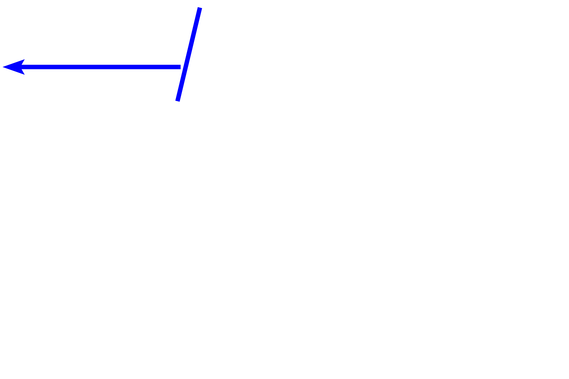
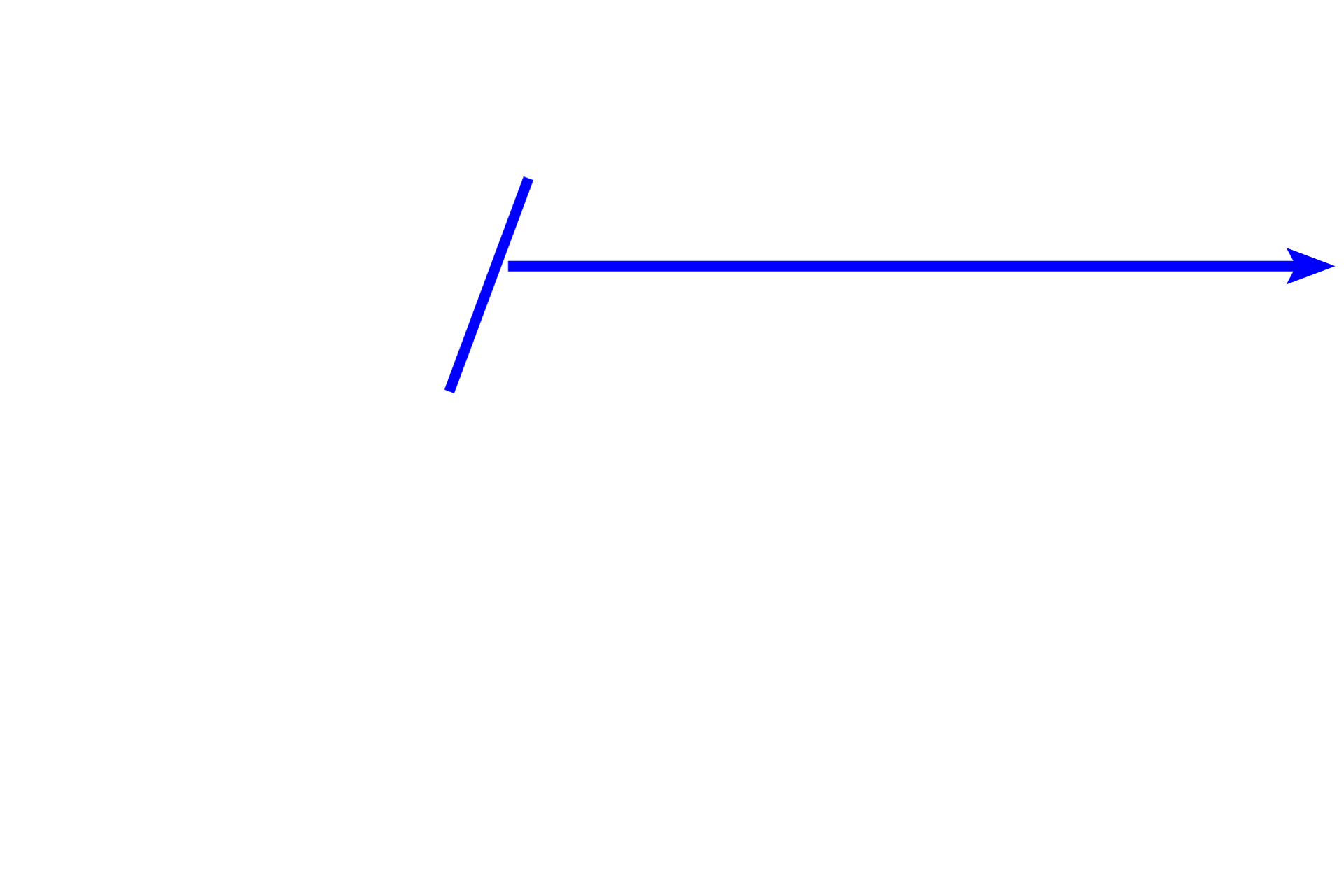
The inner circular layer of the muscularis externa thickens terminally forming the internal anal sphincter (oval). The outer longitudinal layer of the muscularis externa (arrows) decreases and blends with the adventitia.

External anal sphincter >
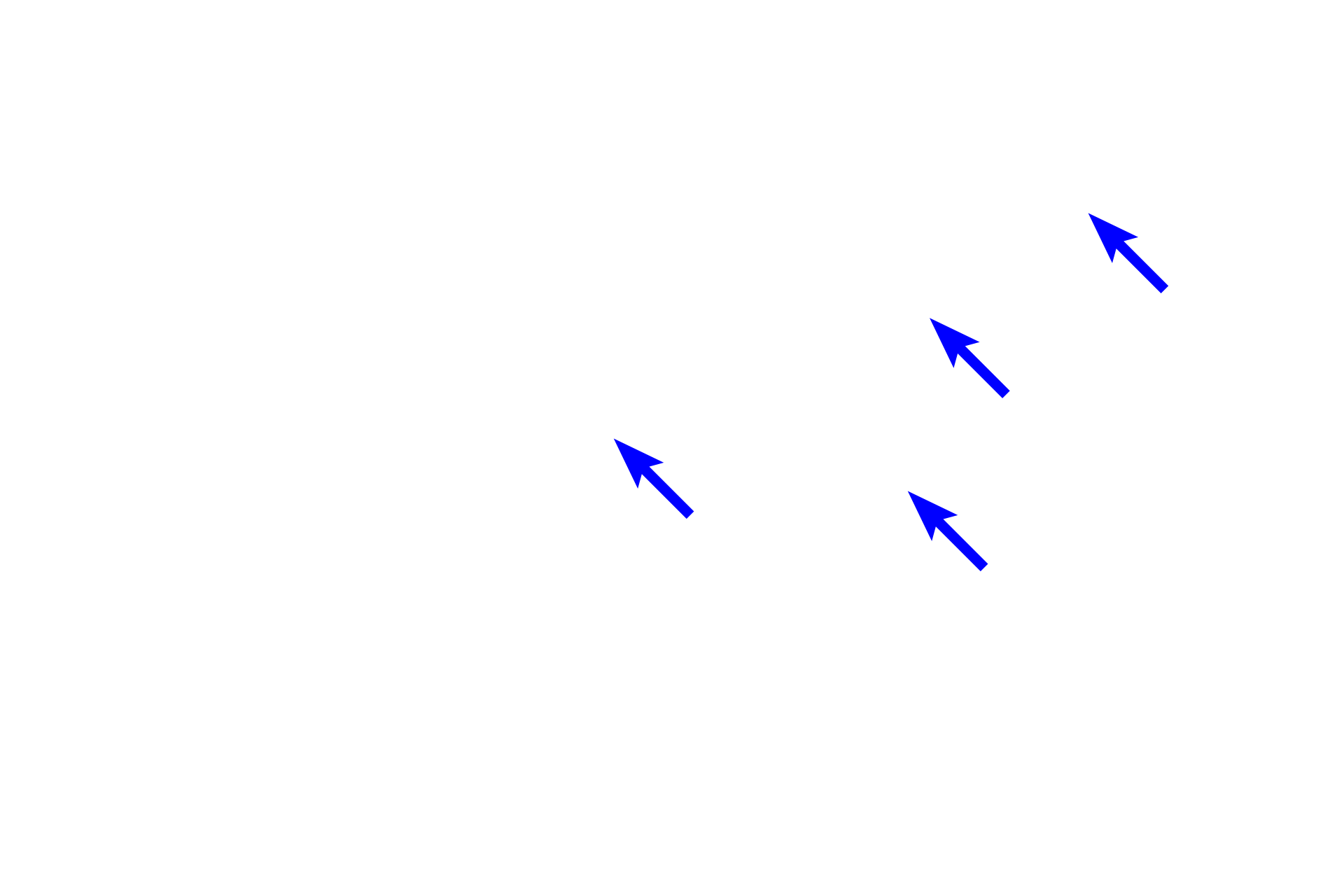
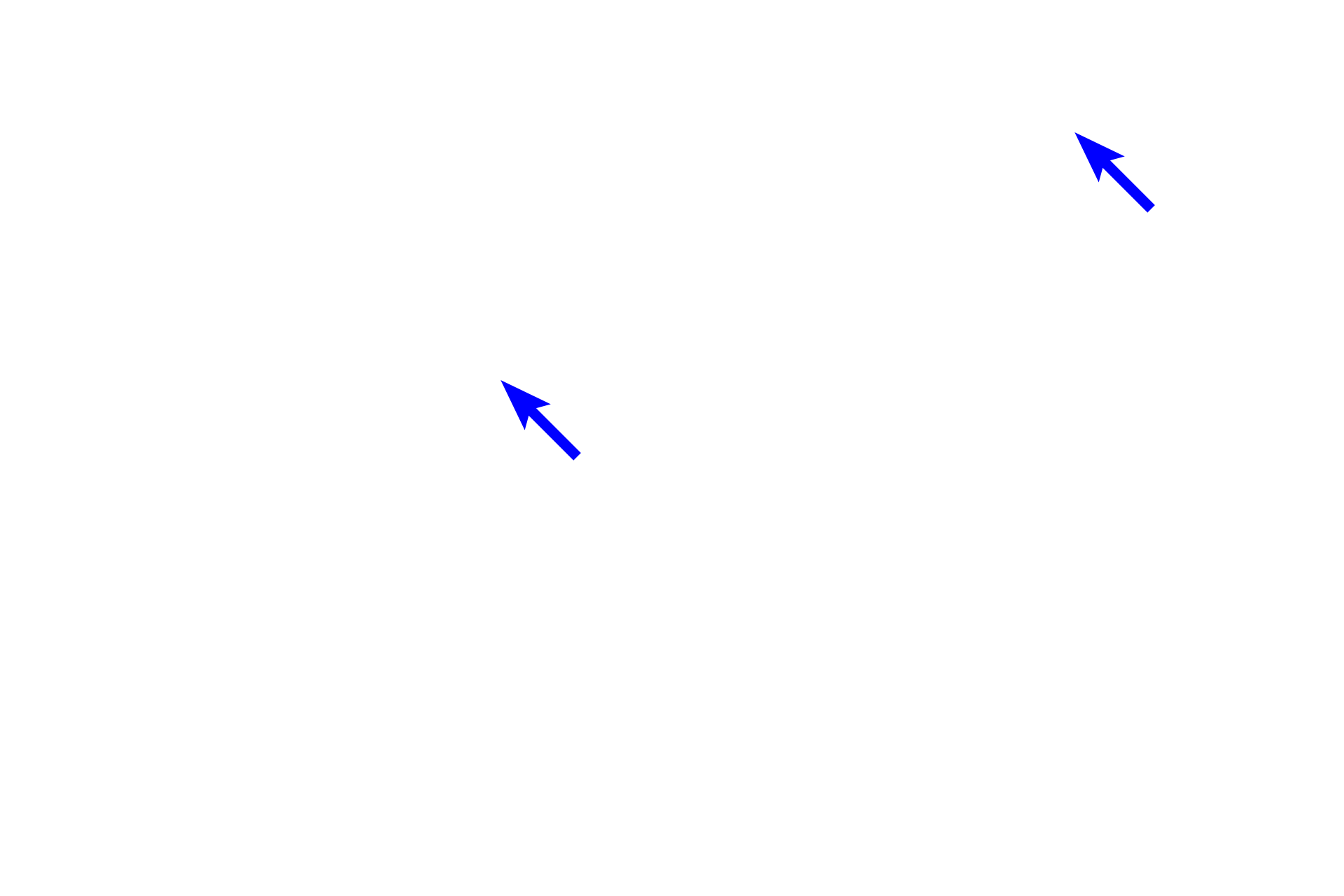
The external anal sphincter is composed of skeletal muscle and consists of deep, superficial and subcutaneous regions. The superficial division (yellow arrows) and the subcutaneous division (blue arrows) are visible in this section.

Next image >
This region is shown in the next image at higher magnification.
Image source >
This images was taken of a slide on the Virtual Microscopy Laboratory website (www.histologyguide.com).