
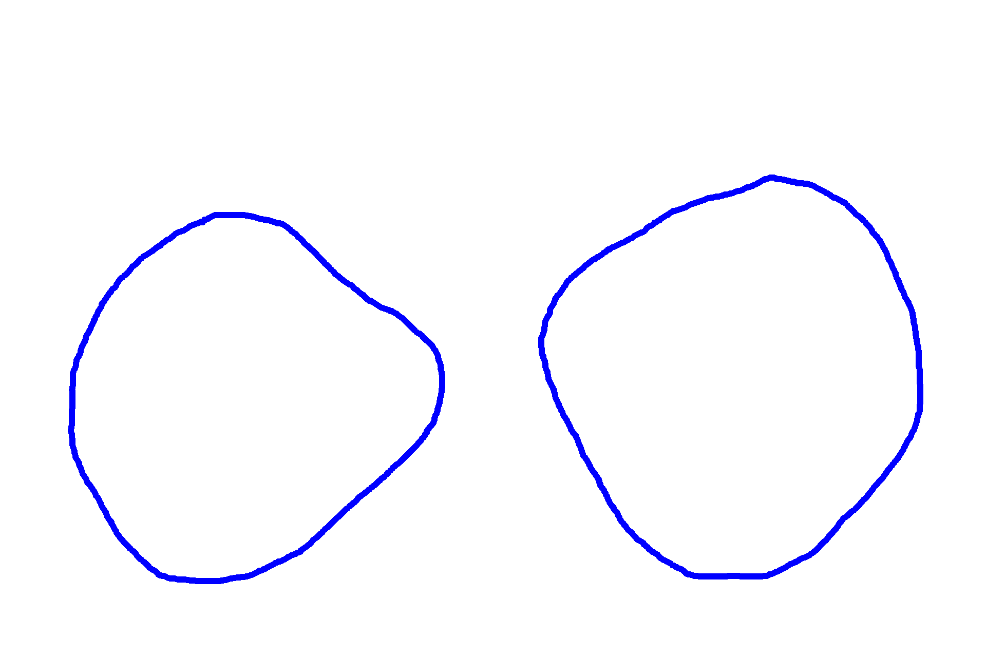
Cap stage of tooth development (early)
These images show early cap stages from opposite sides of the developing oral cavity. During the cap stage of tooth development, cells become identifiable as a tooth germ by the process of histodifferentiation. The cells on the lateral aspects of the original bud begin to rapidly divide, resulting a cup shape, consisting of an inner enamel epithelium and an outer enamel epithelium. The ectomesenchyme encloses by the cup forms the papilla; the surrounding ectomesenchyme forms the dental follicle.
Dental lamina >
The dental lamina connects the developing tooth germ with the oral ectoderm.
Tooth germ >
The tooth germ, first identifiable at the cap stage, is the collection of cells that develop into the tooth. The tooth germ consists of all identifiable ectoderm and ectomesenchyme tissue, including the inner enamel epithelium and outer enamel epithelium (ectoderm) as well as the dental papillae and the dental follicle (ectomesenchyme).

- Enamel organ >
The enamel organ develops from the dental lamina during the cap stage of tooth development. It consists of an inner enamel epithelium, an outer enamel epithelium and a central region containing a stellate reticulum.
- Inner enamel epithelium >
The inner enamel epithelium forms the concave portion of the enamel organ. It consist of columnar cells that differentiate into enamel-forming ameloblasts.
- Outer enamel epithelium >
The outer enamel epithelium serves protective and supportive roles for the tooth germ.
- Cervical loop >
The cervical loop is the site where the inner and outer enamel epithelia meet. It will elongate during tooth development to form Hertwig’s epithelial root sheath.

- Stellate reticulum >
The stellate network supports the inner enamel epithelium and consists of star-shaped cells derived from the epithelium. Their morphology results from secretion of glycosaminoglycans and the resultant attraction of the water into the enamel organ.

Dental papilla >
The dental papilla is formed of ectomesenchymal cells beneath the enamel organ and will form the majority of the dental pulp.
Dental follicle >
The dental follicle, also called the dental sac, surrounds the enamel organ and dental papilla in the apical regions of the tooth germ. The follicle differentiates into cementoblasts that deposit cementum, fibroblasts that deposit collagen fibers as part of the periodontal ligament, and osteoblasts that deposit alveolar bone.

Successional lamina >
Successional lamina develop as an outgrowth from the original dental lamina and form the permanent teeth.