
Developing oral cavity
This image shows a section of the fetal face between 6-8 weeks of gestation, the time period when tooth development begins. Initially, the ectoderm above the primordial mandible and maxilla thickens and extends two horseshoe-shaped laminar evaginations into the underlying ectomesenchyme: a medial dental lamina, which forms the teeth; and a lateral vestibular lamina that forms the vestibule of the oral cavity. 10x
Oral cavity
This image shows a section of the fetal face between 6-8 weeks of gestation, the time period when tooth development begins. Initially, the ectoderm above the primordial mandible and maxilla thickens and extends two horseshoe-shaped laminar evaginations into the underlying ectomesenchyme: a medial dental lamina, which forms the teeth; and a lateral vestibular lamina that forms the vestibule of the oral cavity. 10x

Dental lamina >
The dental lamina forms a horseshoe-shaped epithelial ridge over the developing mandible and maxilla. Localized proliferative activity in the ridge produces a series of epithelial ingrowths (buds) into the ectomesenchyme at sites corresponding to the positions of the future deciduous teeth. The dental lamina connects the developing tooth bud to the epithelium of the oral cavity. From this point, tooth development proceeds in four stages: bud, cap, bell and crown.
- Tooth bud
The dental lamina forms a horseshoe-shaped epithelial ridge over the developing mandible and maxilla. Localized proliferative activity in the ridge produces a series of epithelial ingrowths (buds) into the ectomesenchyme at sites corresponding to the positions of the future deciduous teeth. The dental lamina connects the developing tooth bud to the epithelium of the oral cavity. From this point, tooth development proceeds in four stages: bud, cap, bell and crown.

Tooth germ >
At later stages, the tooth germ develops, consisting of inner and outer enamel epithelia (ectoderm), as well as the dental papillae and dental follicle (ectomesenchyme). The tooth germ is first identifiable at the cap stage of tooth development.

Vestibular lamina >
The vestibular lamina is a thickening of the ectoderm in the lateral aspect of the primordial oral cavity. This infolding develops into the vestibule of the oral cavity.
Tongue
This image shows the developing fetal face between 6-8 weeks of gestation. In the primordial jaw, the ectoderm begins to thicken forming a dental lamina in the medial aspect of the primordial oral cavity and a vestibular lamina in its lateral aspect. 10x
Developing mandible
This image shows the developing fetal face between 6-8 weeks of gestation. In the primordial jaw, the ectoderm begins to thicken forming a dental lamina in the medial aspect of the primordial oral cavity and a vestibular lamina in its lateral aspect. 10x
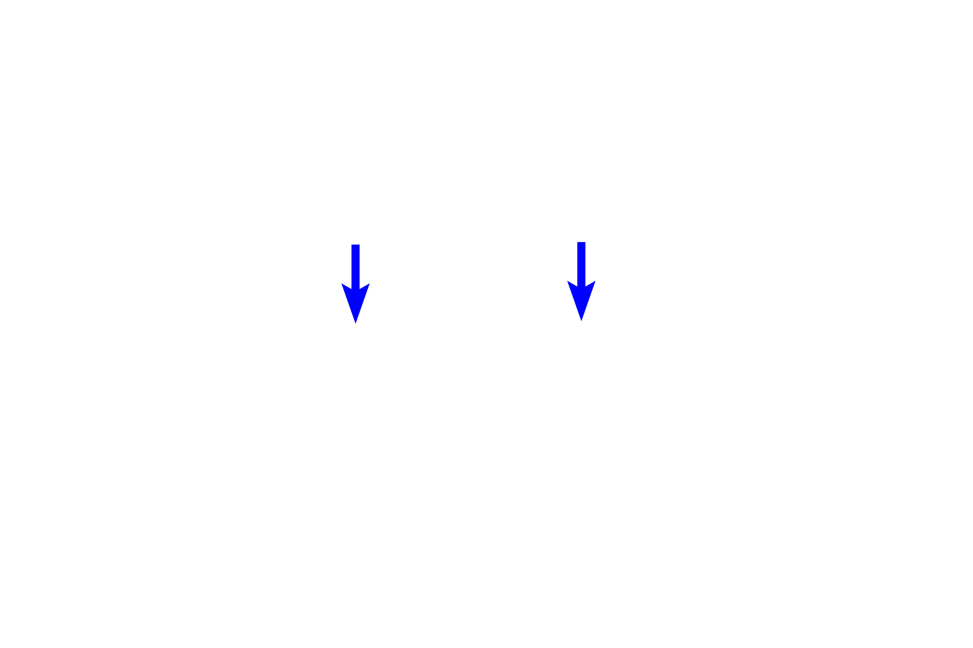
Palate
This image shows the developing fetal face between 6-8 weeks of gestation. In the primordial jaw, the ectoderm begins to thicken forming a dental lamina in the medial aspect of the primordial oral cavity and a vestibular lamina in its lateral aspect. 10x
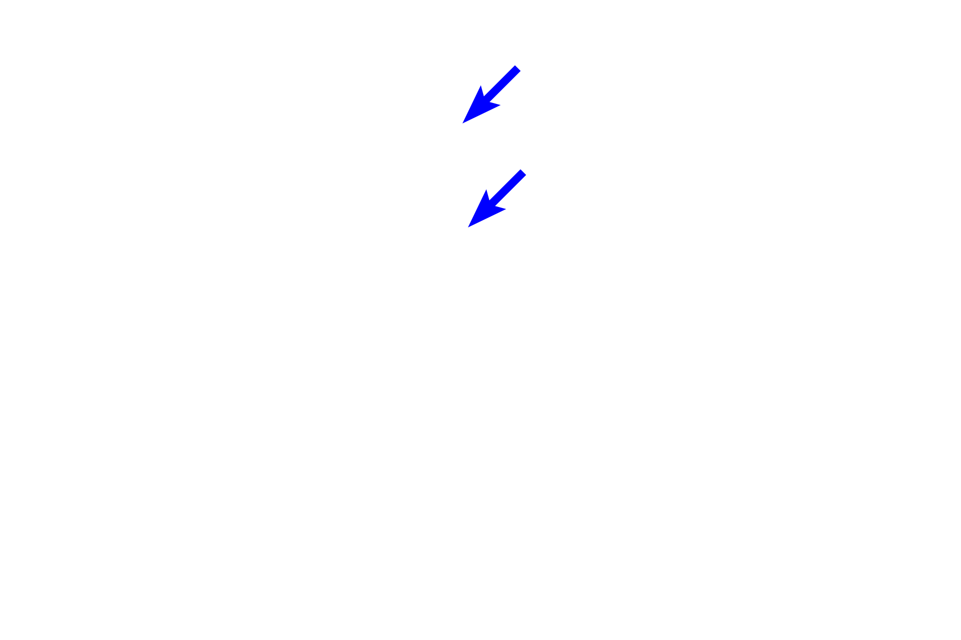
Nasal septum
This image shows the developing fetal face between 6-8 weeks of gestation. In the primordial jaw, the ectoderm begins to thicken forming a dental lamina in the medial aspect of the primordial oral cavity and a vestibular lamina in its lateral aspect. 10x
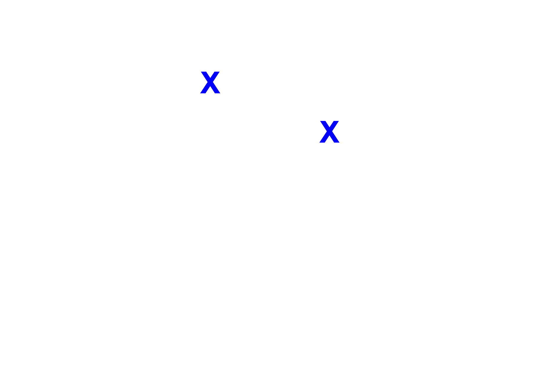
Nasal cavity
This image shows the developing fetal face between 6-8 weeks of gestation. In the primordial jaw, the ectoderm begins to thicken forming a dental lamina in the medial aspect of the primordial oral cavity and a vestibular lamina in its lateral aspect. 10x

Area shown in next image >
This rectangle indicates the area shown at higher magnification in the next image.

Image source >
This image was taken of a slide from the University of South Dakota slide collection.
 PREVIOUS