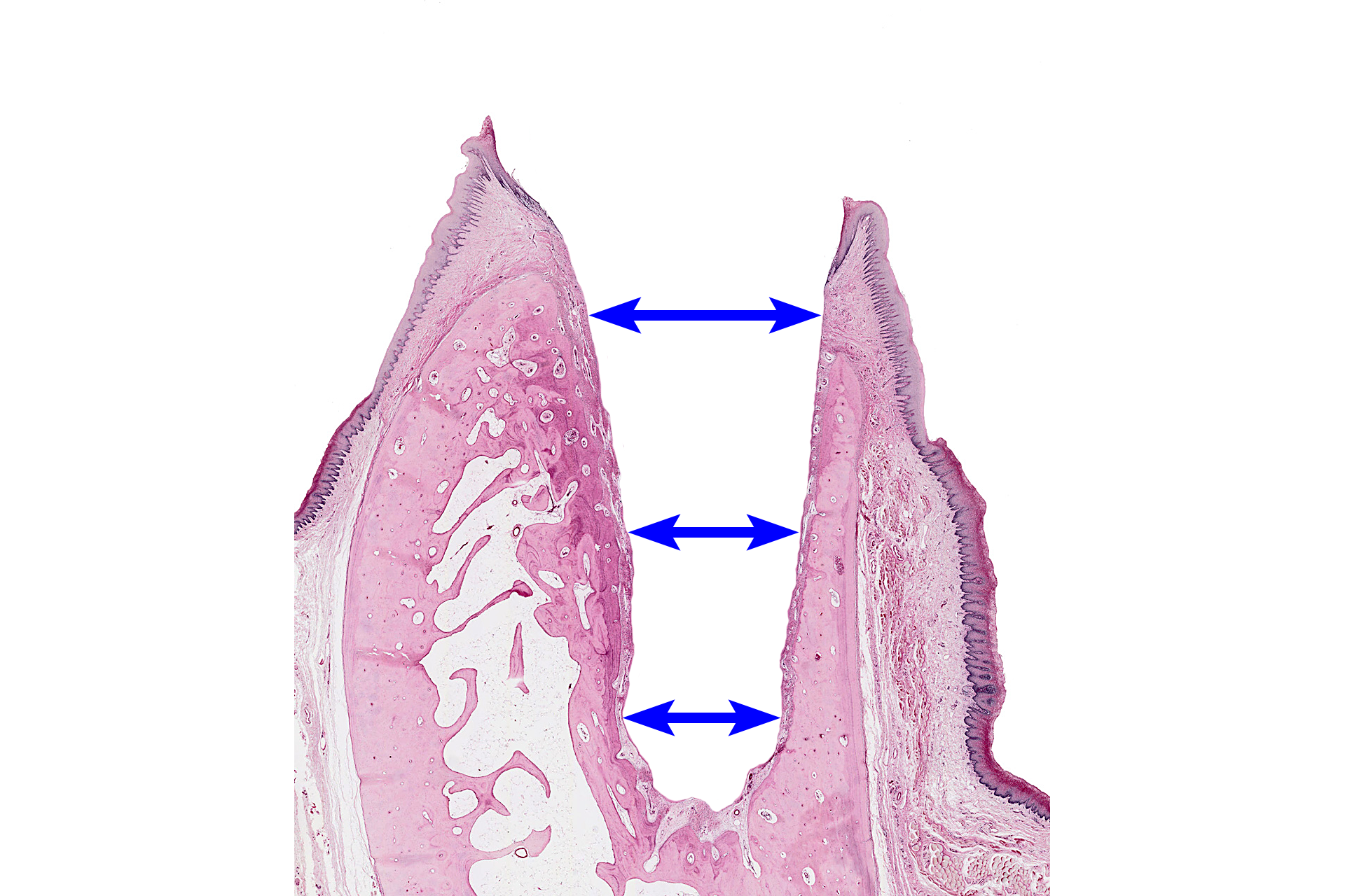
Tooth and dental alveolus
An incisor in its dental alveolus (socket) is shown in this demineralized section. The enamel is not retained as a result of the decalcification process used during tissue preparation. 10x

Dental alveolus >
The dental alveolus is a bony socket that holds the tooth. Dental alveoli are located in the maxilla and mandible. All alveolar bone is differentiated from ectomesenchymal stem cells of the dental follicle. This image was created by digitally removing all the tooth tissues.

- Alveolar bone proper >
Alveolar bone proper forms a thin lining of the alveolus and is located adjacent to the tooth. Periodontal ligaments anchor the tooth in the alveolus by inserting into the alveolar bone proper.

- Spongy supporting alveolar bone >
The majority of the tooth socket is composed of spongy supporting alveolar bone. This bone may be missing in regions where the tooth is leaning to one side, as seen on the right side of in this image.

- Cortical plate alveolar bone >
The lingual and buccal surfaces of the alveolus are covered by cortical plate alveolar bone, a layer of compact bone that is thicker than alveolar bone proper.

- Alveolar crest bone >
The region of compact bone where alveolar bone proper and cortical plate bone meet is alveolar crest bone.

Tooth >
Teeth are located in bony dental alveoli as illustrated by this incisor.

- Crown >
The crown of the tooth extends above the gum line and is covered by enamel. Enamel is not retained in this section as a result of the decalcification process used during tissue preparation. The position of the enamel in indicated by the blue shaded region.

- Neck (cervix) >
The neck of the tooth is a narrow region where the enamel of the crown meets the cememtum. It includes the dentino-enamel-cementum junction.

- Root >
The root of the tooth occupies the dental alveolus to which it is attached by the periodontal ligament.

- Enamel >
Enamel, consisting of about 96% calcium phosphate (hydroxyapatite), is the most mineralized tissue in the human body. It is not retained in this section as a result of the decalcification process used during tissue preparation.

- Dentin >
Dentin is the first hard tissue deposited during tooth development. It makes up the bulk of the hard tissue of the tooth and is 70% mineralized. Dentin is deposited by odontoblasts that reside in the pulp cavity throughout life.

- Pulp cavity >
The pulp cavity is the space within the tooth and contains dental pulp. The cavity conforms to the general shape of the tooth and reduces with size with age.

-- Pulp >
Pulp fills the pulp cavity and consists of loose connective tissue containing a variety of cell types, including ectomesenchymal stem cells, odontoblasts, and immune cells. The pulp also contains blood vessels and nerves that enter at the apical foramen of each root. The pulp is continuous with the connective tissue of the periodontal ligament at the apical foramen (blue arrow). Pulp is the only nonmineralized tissue in the tooth.

- Cementum >
Cementum is the calcified tissue layer on the surface of the root. It is less mineralized than dentin, roughly 50% organic matrix (cementoid) and 50% hydroxyapatite; similar to bone. Cementum functions to attach the tooth to the tooth socket by anchoring the periodontal ligament. Cementum also seals the ends of the dentin tubules and allows for repair at the tooth root.

-- Acellular cementum
Acellular cementum is a thin layer of mineralized tissue on the surface of the cervical two-thirds of the root. It is the first cementum and does not contain cells.

-- Cellular cementum
Cellular cementum contains cells (cementocytes) and is present on the apical one-third of the root. It is thicker than acellular cementum and produced throughout life in response to force or trauma.

Periodontal ligament >
The periodontal ligament anchors the tooth in the socket, protects the tooth by providing a shock absorber, and provides vascular supply and nutrients to the tooth. It also contains inflammatory cells, macrophages and lymphocytes, to defend the tooth against infections that result in periodontal disease. The periodontal ligament develops from the ectomesenchymal stem cells of the dental follicle during root formation.

Gingiva >
The gingiva (commonly called the gums) is composed of the epithelia and connective tissue that surround and support the tooth. The outer portion of the gingiva is formed by the masticatory mucosa, composed of stratified squamous moist orthokeratinized or parakeratinized epithelia and an underlying lamina propria. The highly interdigitated epithelial ridges and long connective tissue papillae firmly attach the epithelium to the lamina propria.

Lining mucosa and submucosa >
Lining mucosa (rectangles) consists of a stratified squamous non-keratinized (moist) epithelium along with its lamina propria. It covers alveolar regions adjacent to the gingiva as well as the remainder of the oral cavity except for the hard palate and the dorsal surface of the tongue. The papillae of the lamina propria are shorter and less numerous than those of the masticatory mucosa of the gingiva. A submucosa (arrows) is present beneath the lining mucosa and contains loose connective tissue, blood vessels, nerves, and minor salivary glands.

Image source >
This image was taken of a slide from the University of Michigan slide collection.