Overview of the cardiovascular system
The major subdivisions of the cardiovascular system include blood (a specialized CT), the heart (left image) which pumps the blood, and blood vessels (right images) that transport the blood. The heart and blood vessels are both composed of three tunics, each of which is similar in composition to, and continuous with, its counterpart in the other subdivision. 10X, 100X

Heart components >
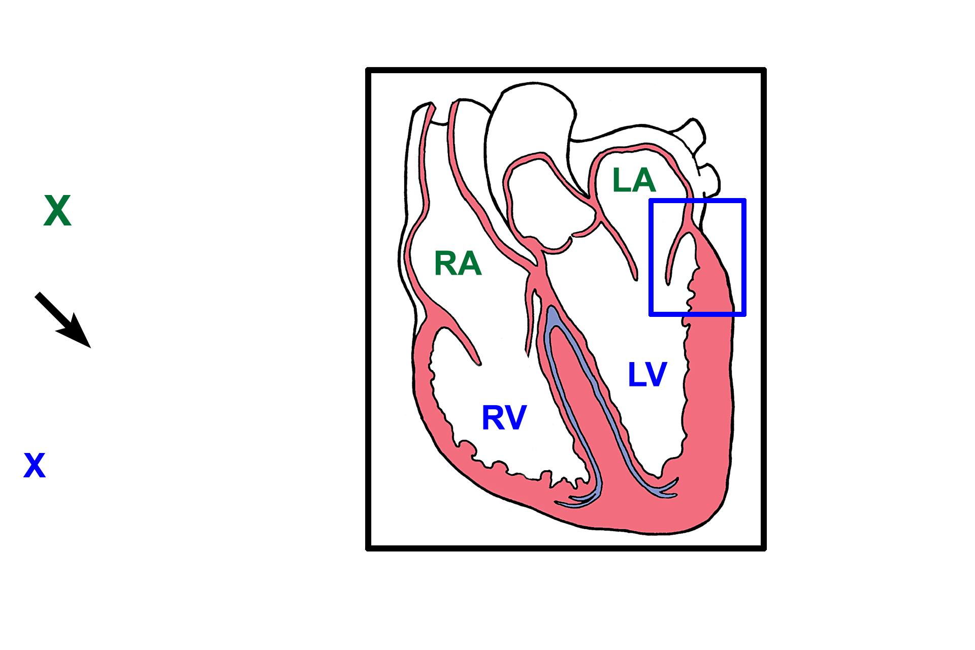
This section of the heart was taken from an area similar to that outlined by the blue rectangle in the illustration. The left atrium (green X) is the upper chamber; the left ventricle (blue X) is the lower chamber; a flap of the atrioventricular valve (arrow) separates the two chambers.

Inner tunic >
The innermost tunic of either subdivision faces the blood and is called the endocardium in the heart and the tunica intima in blood vessels. In both subdivisions the innermost tunic is composed of a specialized simple squamous epithelium, called endothelium, and its underlying connective tissue.

Middle tunic >
The middle tunic of the heart is called the myocardium and is composed of cardiac muscle. In blood vessels the middle tunic is the tunica media and is composed of smooth muscle or smooth muscle plus connective tissue components.

Outer tunic >
The heart is suspended in an internal body cavity (pericardium), so its outer tunic is a serous membrane (epicardium). Like all serous membranes, epicardium is composed of a CT layer covered by a simple squamous epithelium (mesothelium). In blood vessels the outermost tunic is tunica adventitia, a CT layer, although smooth muscle is present in this tunic in large veins.

Blood supply >
The heart and larger blood vessels are so large that they require their own blood supply, which is located in the outermost tunic of both subdivisions. Coronary vessels supply oxygen and nutrients to the heart while vasa vasorum (\”vessels of the vessels\”) are the equivalent blood supply to the larger vessels.