
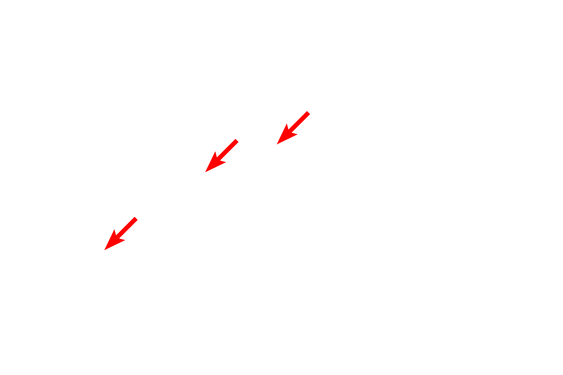
Membranes
This image shows a sample of unmyelinated axons prepared using the freeze-fracture method. In this technique, tissue is frozen and then fractured, which separates membranes into the two leaflets of the lipid bilayer. This unique process reveals membrane structure, particularly the membrane proteins which appear as bumps embedded in the lipid leaflet. In this image, the axons are arranged horizontally. 100,000x
Membrane proteins
This image shows a sample of unmyelinated axons prepared using the freeze-fracture method. In this technique, tissue is frozen and then fractured, which separates membranes into the two leaflets of the lipid bilayer. This unique process reveals membrane structure, particularly the membrane proteins which appear as bumps embedded in the lipid leaflet. In this image, the axons are arranged horizontally. 100,000x
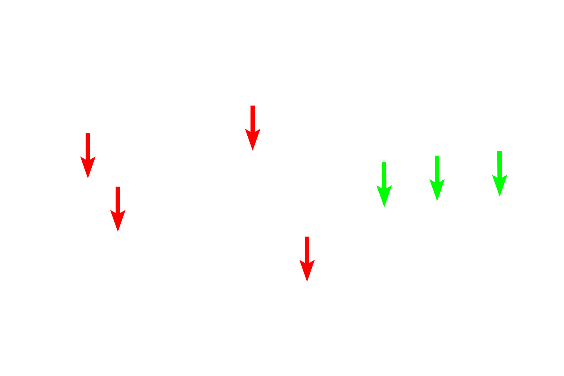
Lipid leaflet >
The green arrows indicate the fractured margin of a leaflet.