
Golgi apparatus
The Golgi consists of stacks of smooth, flattened membranous sacs (cisternae), usually located near the nucleus. Newly synthesized proteins are transferred from the RER by transport vesicles, which fuse with the forming, or cis, face of the Golgi. From there, proteins move through the mid-Golgi where they are modified, and eventually exit in vesicles from the maturing, or trans, face. The resultant collection of vesicles, along with a network of microtubules, form the trans-Golgi network. The microtubules provide the tracks along which the vesicles are transported throughout the cell. 25,000x
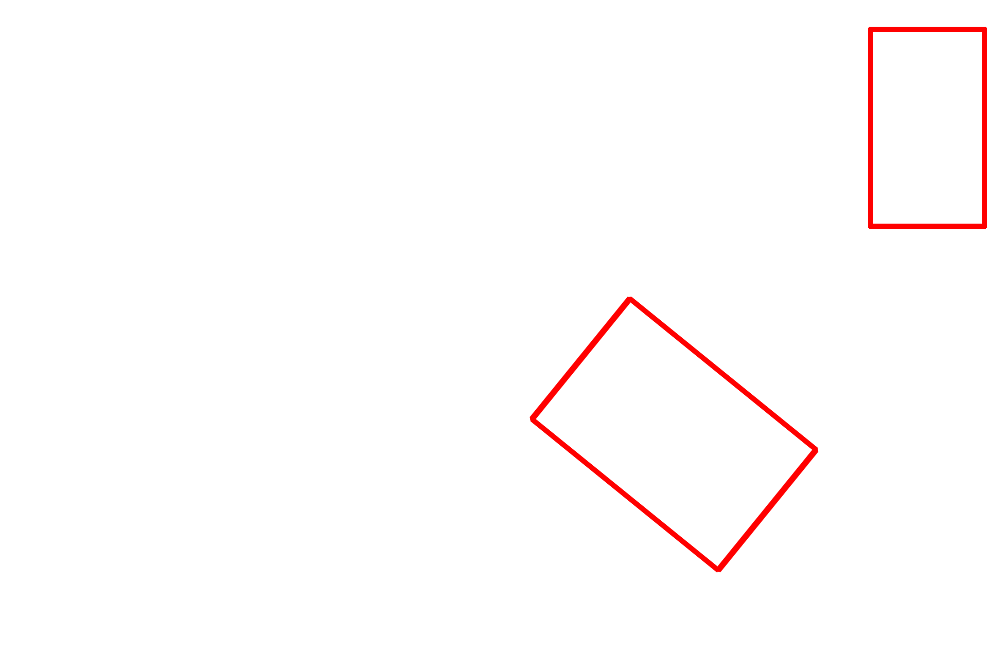
Golgi complexes
The Golgi consists of stacks of smooth, flattened membranous sacs (cisternae), usually located near the nucleus. Newly synthesized proteins are transferred from the RER by transport vesicles, which fuse with the forming, or cis, face of the Golgi. From there, proteins move through the mid-Golgi where they are modified, and eventually exit in vesicles from the maturing, or trans, face. The resultant collection of vesicles, along with a network of microtubules, form the trans-Golgi network. The microtubules provide the tracks along which the vesicles are transported throughout the cell. 25,000x

- Cis face
The Golgi consists of stacks of smooth, flattened membranous sacs (cisternae), usually located near the nucleus. Newly synthesized proteins are transferred from the RER by transport vesicles, which fuse with the forming, or cis, face of the Golgi. From there, proteins move through the mid-Golgi where they are modified, and eventually exit in vesicles from the maturing, or trans, face. The resultant collection of vesicles, along with a network of microtubules, form the trans-Golgi network. The microtubules provide the tracks along which the vesicles are transported throughout the cell. 25,000x

- Mid-Golgi region
The Golgi consists of stacks of smooth, flattened membranous sacs (cisternae), usually located near the nucleus. Newly synthesized proteins are transferred from the RER by transport vesicles, which fuse with the forming, or cis, face of the Golgi. From there, proteins move through the mid-Golgi where they are modified, and eventually exit in vesicles from the maturing, or trans, face. The resultant collection of vesicles, along with a network of microtubules, form the trans-Golgi network. The microtubules provide the tracks along which the vesicles are transported throughout the cell. 25,000x

- Trans face
The Golgi consists of stacks of smooth, flattened membranous sacs (cisternae), usually located near the nucleus. Newly synthesized proteins are transferred from the RER by transport vesicles, which fuse with the forming, or cis, face of the Golgi. From there, proteins move through the mid-Golgi where they are modified, and eventually exit in vesicles from the maturing, or trans, face. The resultant collection of vesicles, along with a network of microtubules, form the trans-Golgi network. The microtubules provide the tracks along which the vesicles are transported throughout the cell. 25,000x

- Trans Golgi network
The Golgi consists of stacks of smooth, flattened membranous sacs (cisternae), usually located near the nucleus. Newly synthesized proteins are transferred from the RER by transport vesicles, which fuse with the forming, or cis, face of the Golgi. From there, proteins move through the mid-Golgi where they are modified, and eventually exit in vesicles from the maturing, or trans, face. The resultant collection of vesicles, along with a network of microtubules, form the trans-Golgi network. The microtubules provide the tracks along which the vesicles are transported throughout the cell. 25,000x

Nucleus
The Golgi consists of stacks of smooth, flattened membranous sacs (cisternae), usually located near the nucleus. Newly synthesized proteins are transferred from the RER by transport vesicles, which fuse with the forming, or cis, face of the Golgi. From there, proteins move through the mid-Golgi where they are modified, and eventually exit in vesicles from the maturing, or trans, face. The resultant collection of vesicles, along with a network of microtubules, form the trans-Golgi network. The microtubules provide the tracks along which the vesicles are transported throughout the cell. 25,000x

Mitochondrion
The Golgi consists of stacks of smooth, flattened membranous sacs (cisternae), usually located near the nucleus. Newly synthesized proteins are transferred from the RER by transport vesicles, which fuse with the forming, or cis, face of the Golgi. From there, proteins move through the mid-Golgi where they are modified, and eventually exit in vesicles from the maturing, or trans, face. The resultant collection of vesicles, along with a network of microtubules, form the trans-Golgi network. The microtubules provide the tracks along which the vesicles are transported throughout the cell. 25,000x

Lysosome
The Golgi consists of stacks of smooth, flattened membranous sacs (cisternae), usually located near the nucleus. Newly synthesized proteins are transferred from the RER by transport vesicles, which fuse with the forming, or cis, face of the Golgi. From there, proteins move through the mid-Golgi where they are modified, and eventually exit in vesicles from the maturing, or trans, face. The resultant collection of vesicles, along with a network of microtubules, form the trans-Golgi network. The microtubules provide the tracks along which the vesicles are transported throughout the cell. 25,000x
Polysomes
The Golgi consists of stacks of smooth, flattened membranous sacs (cisternae), usually located near the nucleus. Newly synthesized proteins are transferred from the RER by transport vesicles, which fuse with the forming, or cis, face of the Golgi. From there, proteins move through the mid-Golgi where they are modified, and eventually exit in vesicles from the maturing, or trans, face. The resultant collection of vesicles, along with a network of microtubules, form the trans-Golgi network. The microtubules provide the tracks along which the vesicles are transported throughout the cell. 25,000x
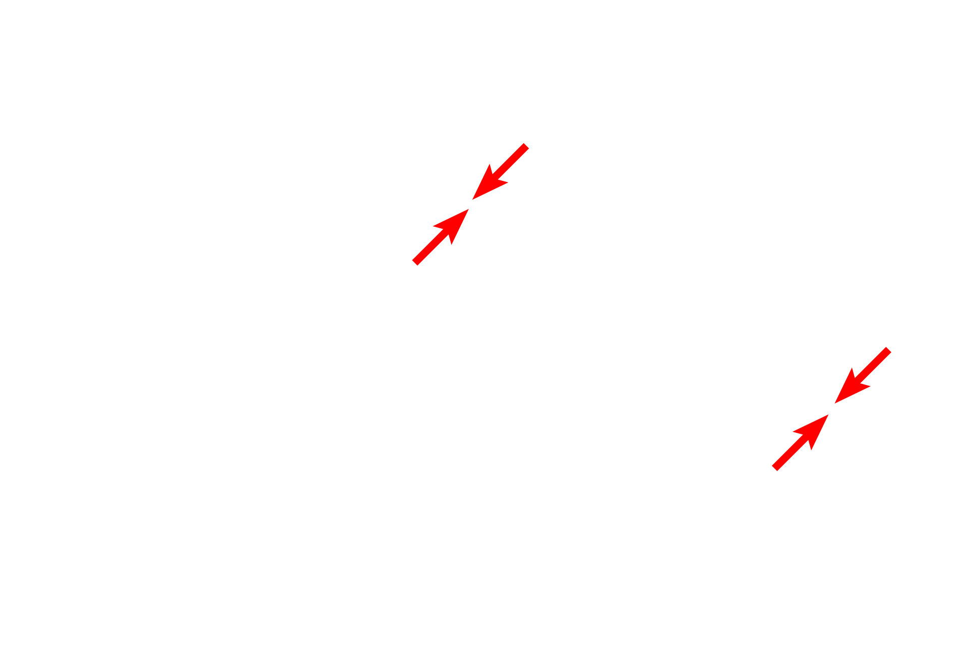
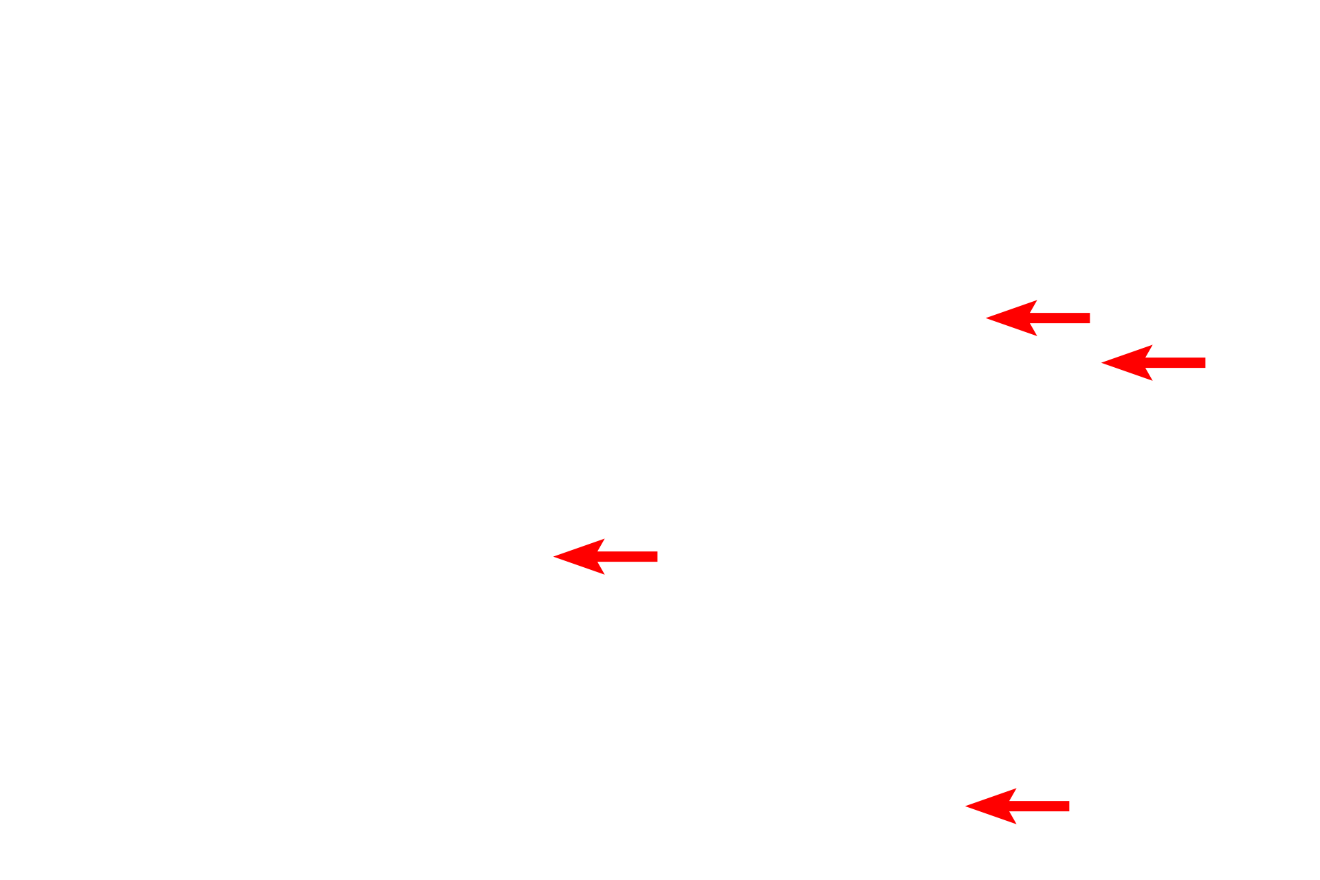
Plasma membranes >
The plasma membranes of the cells can be seen running diagonally through the micrograph.
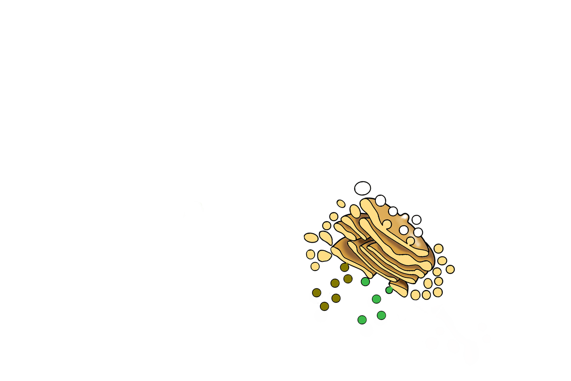
Illustration of the Golgi apparatus >
This overlay drawing of the Golgi apparatus shows the relationship of its components.