
Glycogen
These images compare the appearance of glycogen in hepatocytes by light microscopy (left, PAS stain) and electron microscopy (right). Individual glycogen granules, which measure 15 to 40 nm in diameter, are too small to be resolved by light microscopy. However, aggregates of these granules, which measure up to 300 nm in diameter, can be visualized. By electron microscopy, aggregates and their individual granules are clearly evident. 1000x (l); 7500x (r)
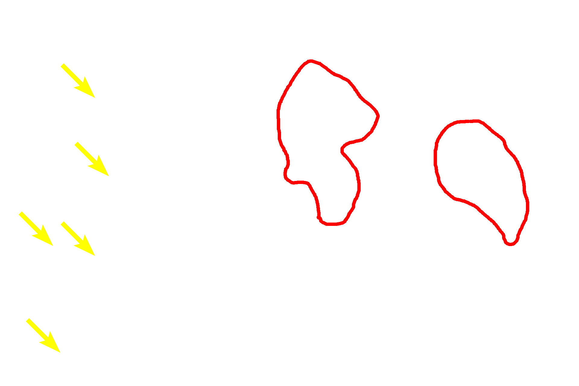
Glycogen aggregates
These images compare the appearance of glycogen in hepatocytes by light microscopy (left, PAS stain) and electron microscopy (right). Individual glycogen granules, which measure 15 to 40 nm in diameter, are too small to be resolved by light microscopy. However, aggregates of these granules, which measure up to 300 nm in diameter, can be visualized. By electron microscopy, aggregates and their individual granules are clearly evident. 1000x (l); 7500x (r)
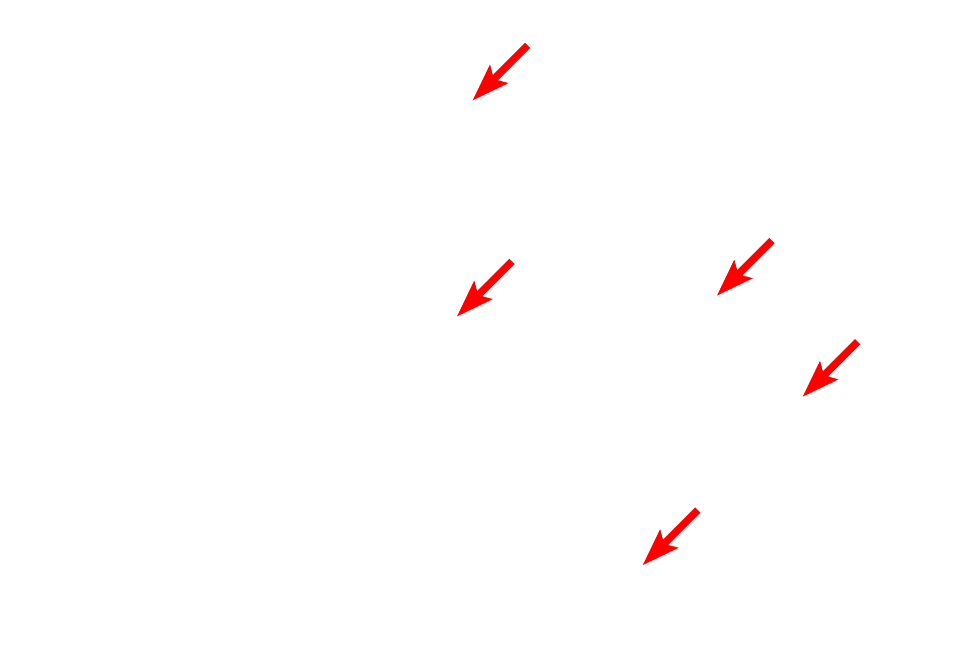
Glycogen granules
These images compare the appearance of glycogen in hepatocytes by light microscopy (left, PAS stain) and electron microscopy (right). Individual glycogen granules, which measure 15 to 40 nm in diameter, are too small to be resolved by light microscopy. However, aggregates of these granules, which measure up to 300 nm in diameter, can be visualized. By electron microscopy, aggregates and their individual granules are clearly evident. 1000x (l); 7500x (r)

Nuclei
These images compare the appearance of glycogen in hepatocytes by light microscopy (left, PAS stain) and electron microscopy (right). Individual glycogen granules, which measure 15 to 40 nm in diameter, are too small to be resolved by light microscopy. However, aggregates of these granules, which measure up to 300 nm in diameter, can be visualized. By electron microscopy, aggregates and their individual granules are clearly evident. 1000x (l); 7500x (r)
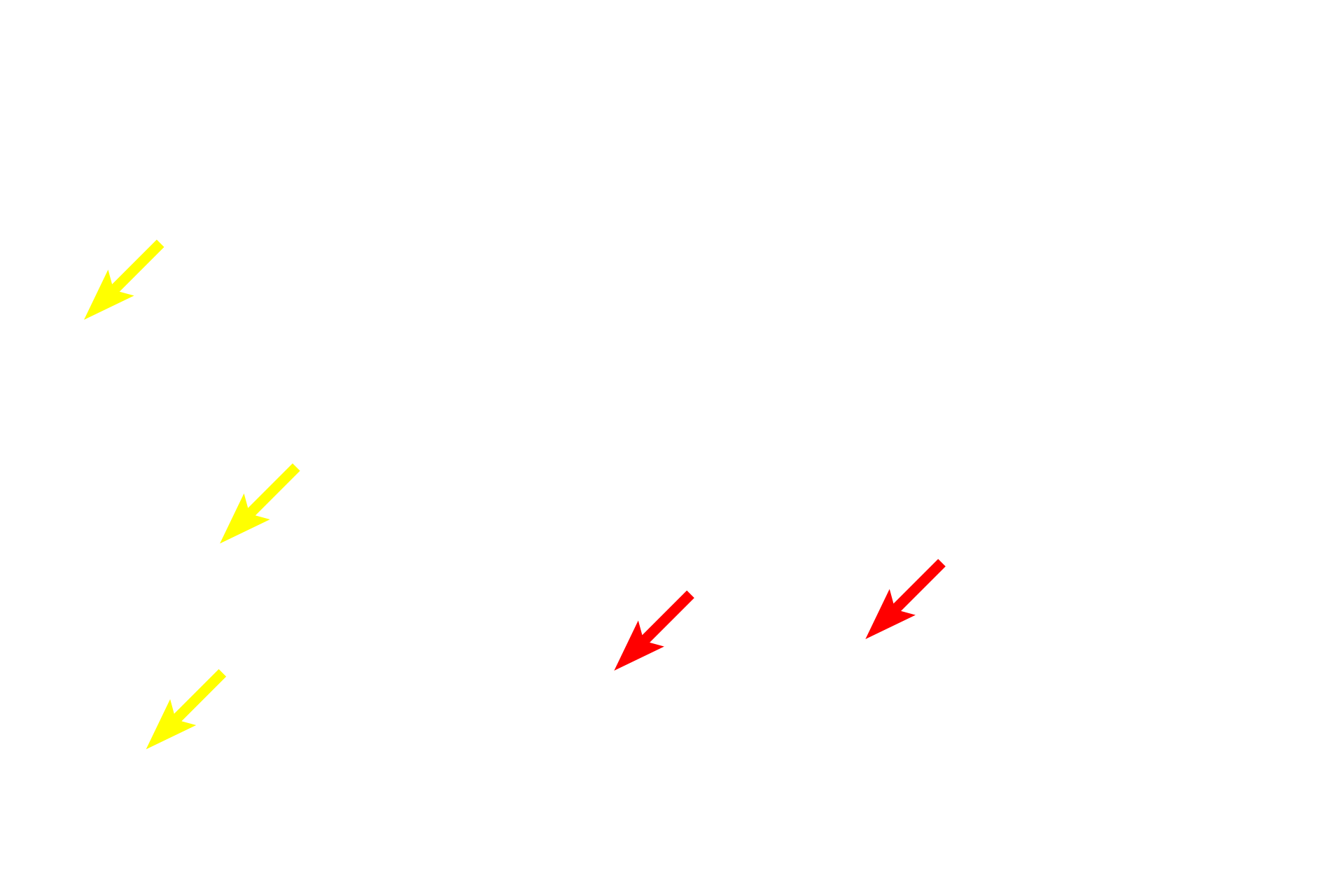
- Nucleoli
These images compare the appearance of glycogen in hepatocytes by light microscopy (left, PAS stain) and electron microscopy (right). Individual glycogen granules, which measure 15 to 40 nm in diameter, are too small to be resolved by light microscopy. However, aggregates of these granules, which measure up to 300 nm in diameter, can be visualized. By electron microscopy, aggregates and their individual granules are clearly evident. 1000x (l); 7500x (r)
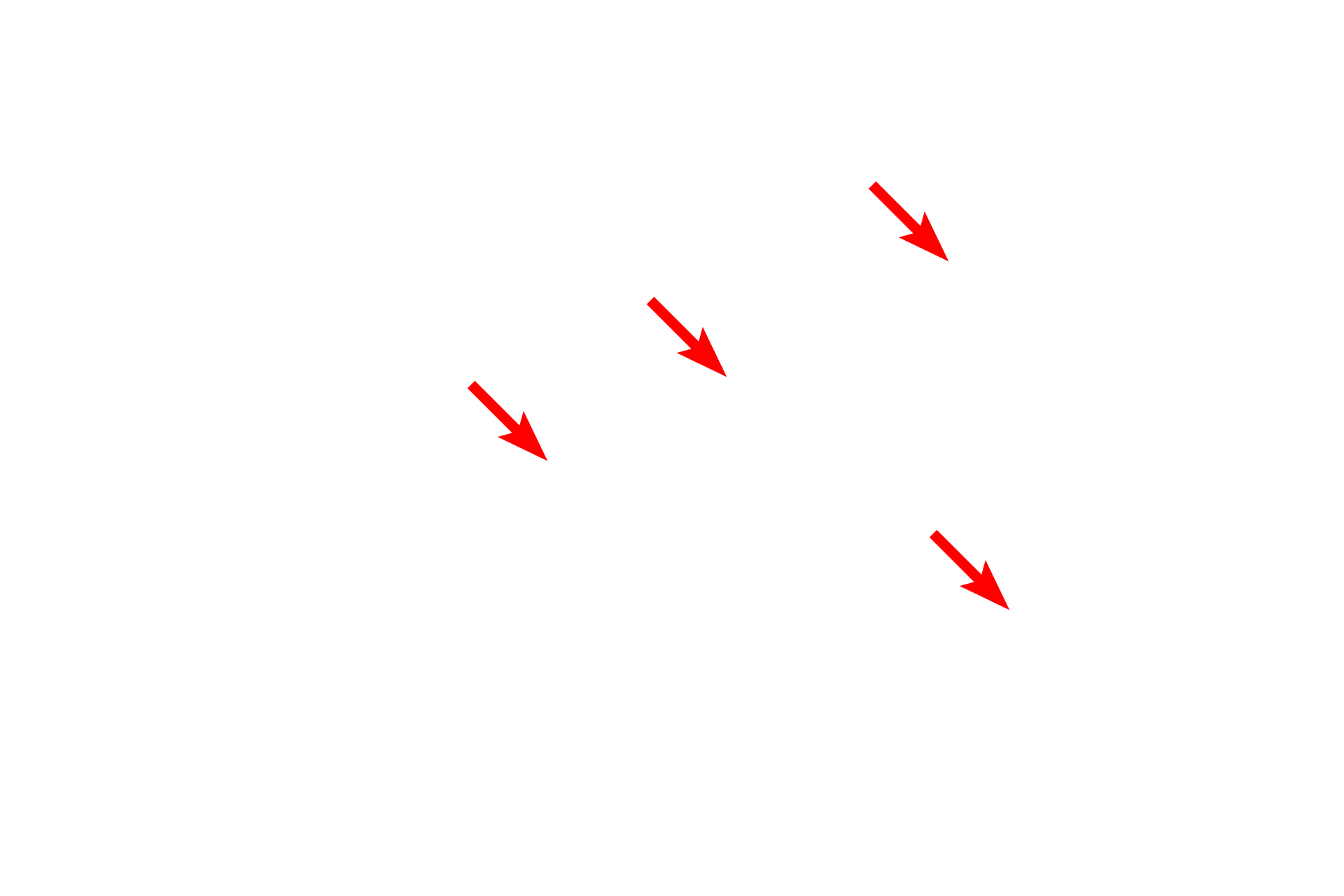
Mitochondria
These images compare the appearance of glycogen in hepatocytes by light microscopy (left, PAS stain) and electron microscopy (right). Individual glycogen granules, which measure 15 to 40 nm in diameter, are too small to be resolved by light microscopy. However, aggregates of these granules, which measure up to 300 nm in diameter, can be visualized. By electron microscopy, aggregates and their individual granules are clearly evident. 1000x (l); 7500x (r)
RER
These images compare the appearance of glycogen in hepatocytes by light microscopy (left, PAS stain) and electron microscopy (right). Individual glycogen granules, which measure 15 to 40 nm in diameter, are too small to be resolved by light microscopy. However, aggregates of these granules, which measure up to 300 nm in diameter, can be visualized. By electron microscopy, aggregates and their individual granules are clearly evident. 1000x (l); 7500x (r)
 PREVIOUS