
Intermediate filaments and microtubules
This electron micrograph shows cross sections of myelinated axons, which possess a mixture of microtubules and intermediate filaments that both run lengthwise in the axon. Brain, 20,000x
Microtubules >
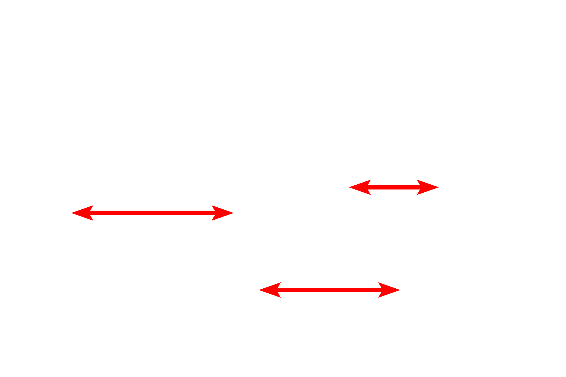
Microtubules are have the largest diameter (20-25 nm) among the cytoskeletal elements and provide the tracks along which materials are transported within cells. Unlike microfilaments and intermediate filaments, which are rod-shaped, microtubules are hollow tubes formed primarily of tubulin proteins. Microtubules also provide some structural support.

Intermediate filaments >
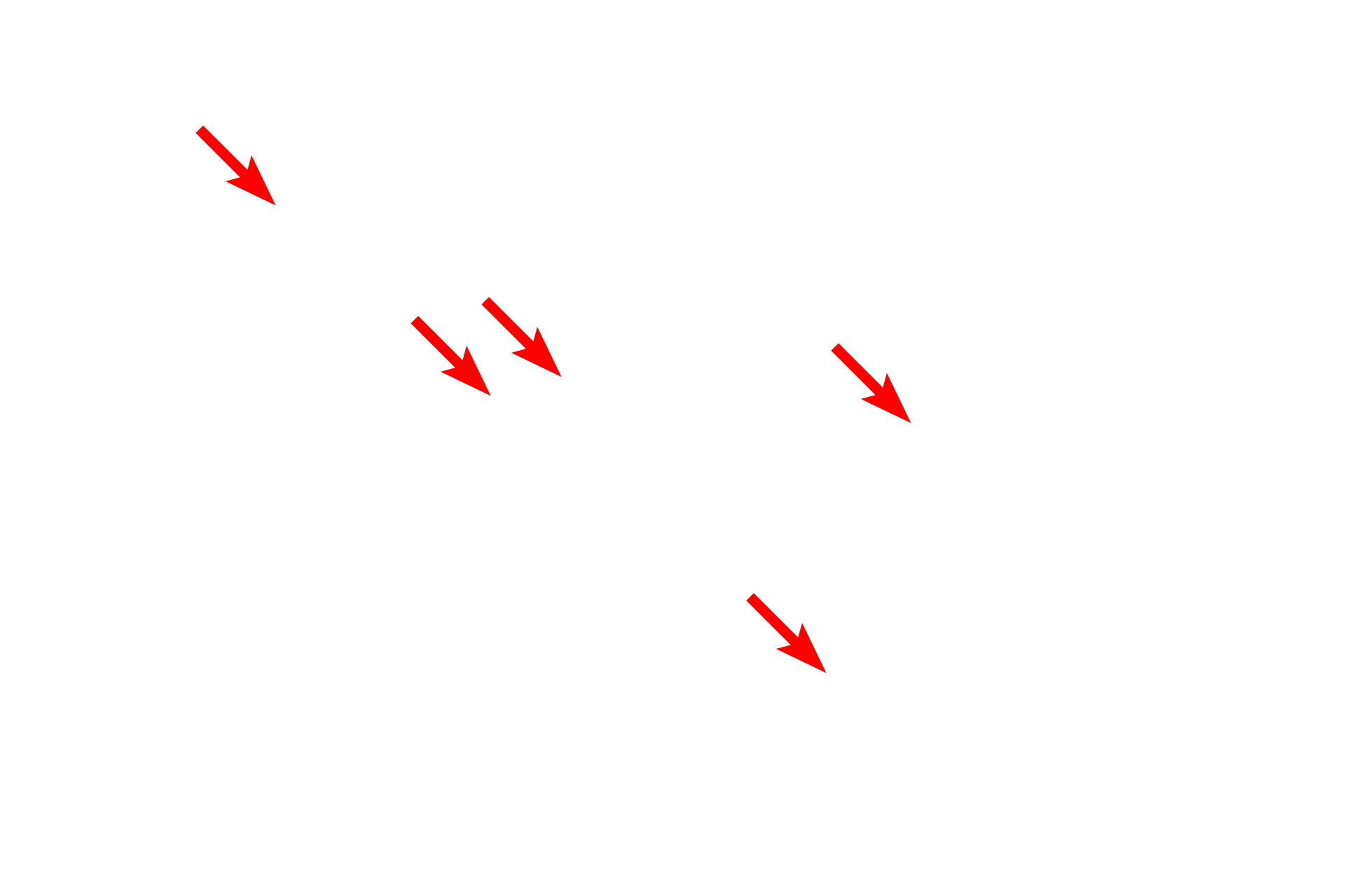
Intermediate filaments occur alongside microtubules in the axon and are called neurofilaments (red arrows). These filaments are rigid and very stable, providing support, particularly in longer, larger axons. Intermediate filaments are also present in supporting astrocytic glial cells; a group of these filaments is outlined in blue. These filaments, called glial filaments, also provide support. Neurofilaments and glial filaments are composed of different proteins.
Axon
This electron micrograph shows cross sections of myelinated axons, which possess a mixture of microtubules and intermediate filaments that both run lengthwise in the axon. Brain, 20,000x

Myelin
This electron micrograph shows cross sections of myelinated axons, which possess a mixture of microtubules and intermediate filaments that both run lengthwise in the axon. Brain, 20,000x

Mitochondria
This electron micrograph shows cross sections of myelinated axons, which possess a mixture of microtubules and intermediate filaments that both run lengthwise in the axon. Brain, 20,000x