
Centrioles
Centrioles are barrel-shaped, non-membrane-bound structures composed of microtubules grouped into nine triplets arranged in a cylinder. Centrioles are most often found in pairs, oriented perpendicularly to each other, forming the diplosome. The diplosome is located in a region of the cell called the centrosome or microtubule organizing center (MTOC). Centrioles are also located individually at the base of cilia and flagella as basal bodies, that organize the microtubules of these structures.
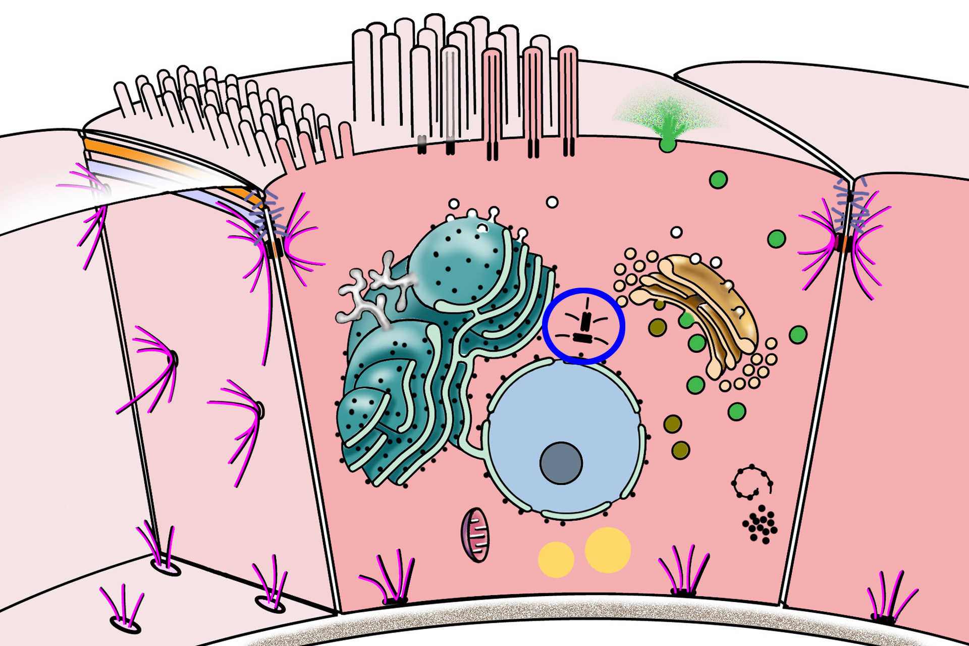
Diplosome and MTOC >
Diplosomes consist of two, perpendicularly-oriented centrioles. The pair is surrounded by dynamic matrix of proteins in a region of the cell, often near the nucleus, called the centrosome or the microtubule-organizing center (MTOC). This region is the primary site for microtubule assembly in interphase cells, extending microtubules for intracellular transport. The MTOC also forms the microtubules of the mitotic spindle during cell division.
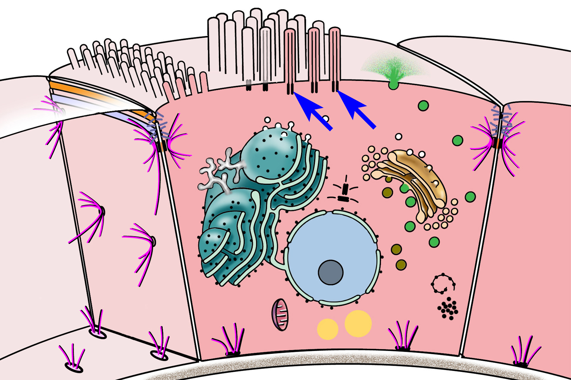
Basal bodies >
Basal bodies are formed by individual centrioles and are located at the bases of cilia and flagella. Basal bodies act as organizing centers for microtubules that extend into each cilium or flagellum. These microtubules form the axoneme, which consists of nine pairs of microtubules (doublets) surrounding two central, individual microtubules (9 + 2 arrangement).
