
Anaphase
This electron micrograph shows a mid-anaphase cell in which the chromosomes have separated and are attached to microtubules of the spindle apparatus. Neither the nuclear envelope nor nucleolus is present. While the nuclear envelope fragments into vesicles, remnants of the RER remain intact. 5000x
Chromosomes
This electron micrograph shows a mid-anaphase cell in which the chromosomes have separated and are attached to microtubules of the spindle apparatus. Neither the nuclear envelope nor nucleolus is present. While the nuclear envelope fragments into vesicles, remnants of the RER remain intact. 5000x
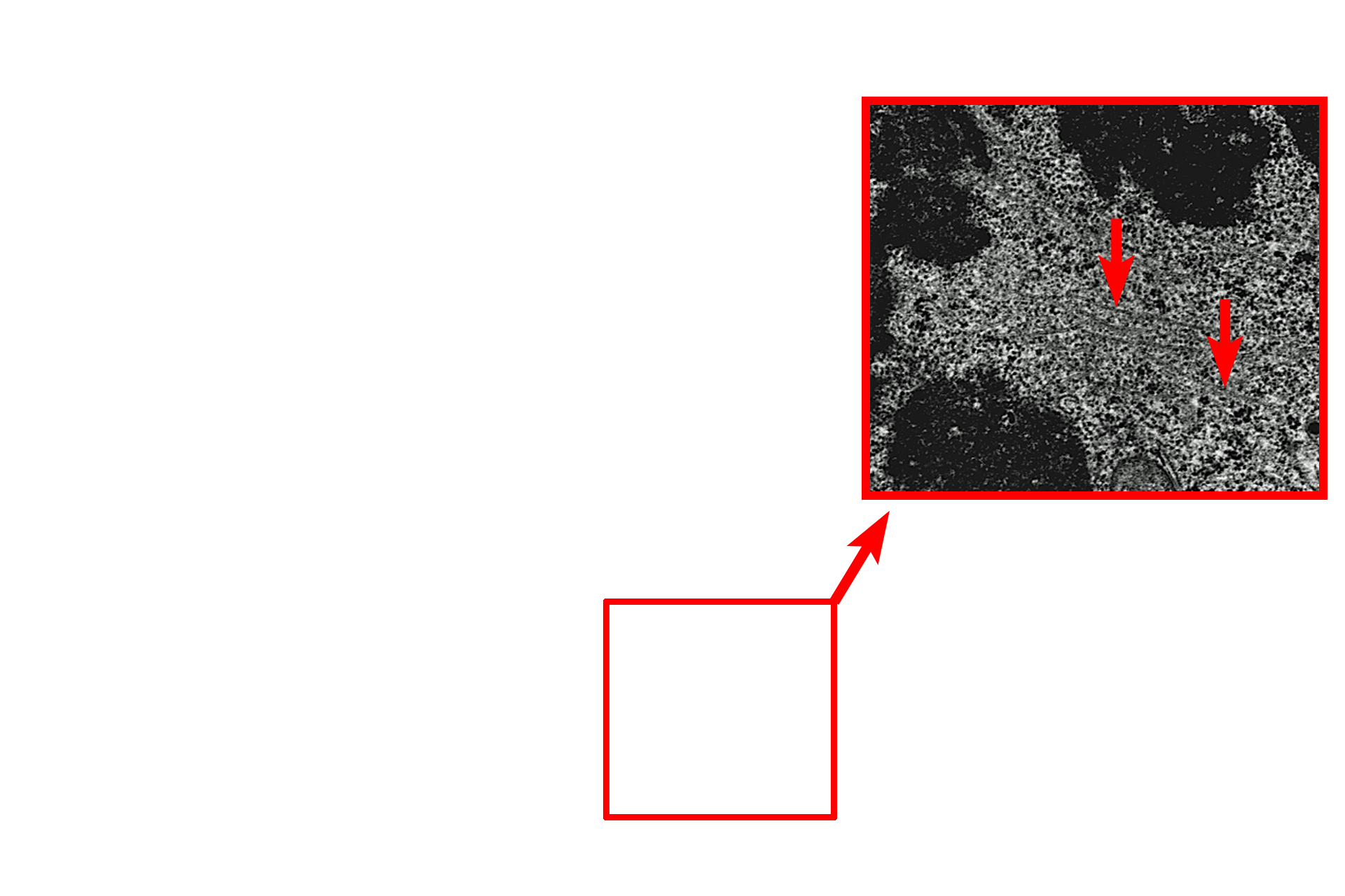
Microtubules >
Kinetochore microtubules are shown at higher magnification in this inset. Kinetochore microtubules extend from the diplosome and attach to the kinetochore of the chromosome.

RER
This electron micrograph shows a mid-anaphase cell in which the chromosomes have separated and are attached to microtubules of the spindle apparatus. Neither the nuclear envelope nor nucleolus is present. While the nuclear envelope fragments into vesicles, remnants of the RER remain intact. 5000x
Mitochondria
This electron micrograph shows a mid-anaphase cell in which the chromosomes have separated and are attached to microtubules of the spindle apparatus. Neither the nuclear envelope nor nucleolus is present. While the nuclear envelope fragments into vesicles, remnants of the RER remain intact. 5000x