
Meiosis II: Anaphase
Anaphase begins abruptly as sister chromatids separate and each is drawn towards an opposite pole of the cell. The movement of the chromatids occurs by the shortening of the spindle fibers and movement of the spindle poles. The end of anaphase is marked by the segregation of a single set of chromosomes to each spindle pole.
Chromosomes >
Sister chromatids are separated from each other following the breakdown of cohesive proteins holding them together at the centromere. Chromatids are then drawn to opposite poles by the pulling force of the kinetochore microtubules. Once separated, chromatids are referred to as chromosomes.
Centrosomes
Sister chromatids are separated from each other following the breakdown of cohesive proteins holding them together at the centromere. Chromatids are then drawn to opposite poles by the pulling force of the kinetochore microtubules. Once separated, chromatids are referred to as chromosomes.
Centromeres
Sister chromatids are separated from each other following the breakdown of cohesive proteins holding them together at the centromere. Chromatids are then drawn to opposite poles by the pulling force of the kinetochore microtubules. Once separated, chromatids are referred to as chromosomes.
Mitotic spindles >
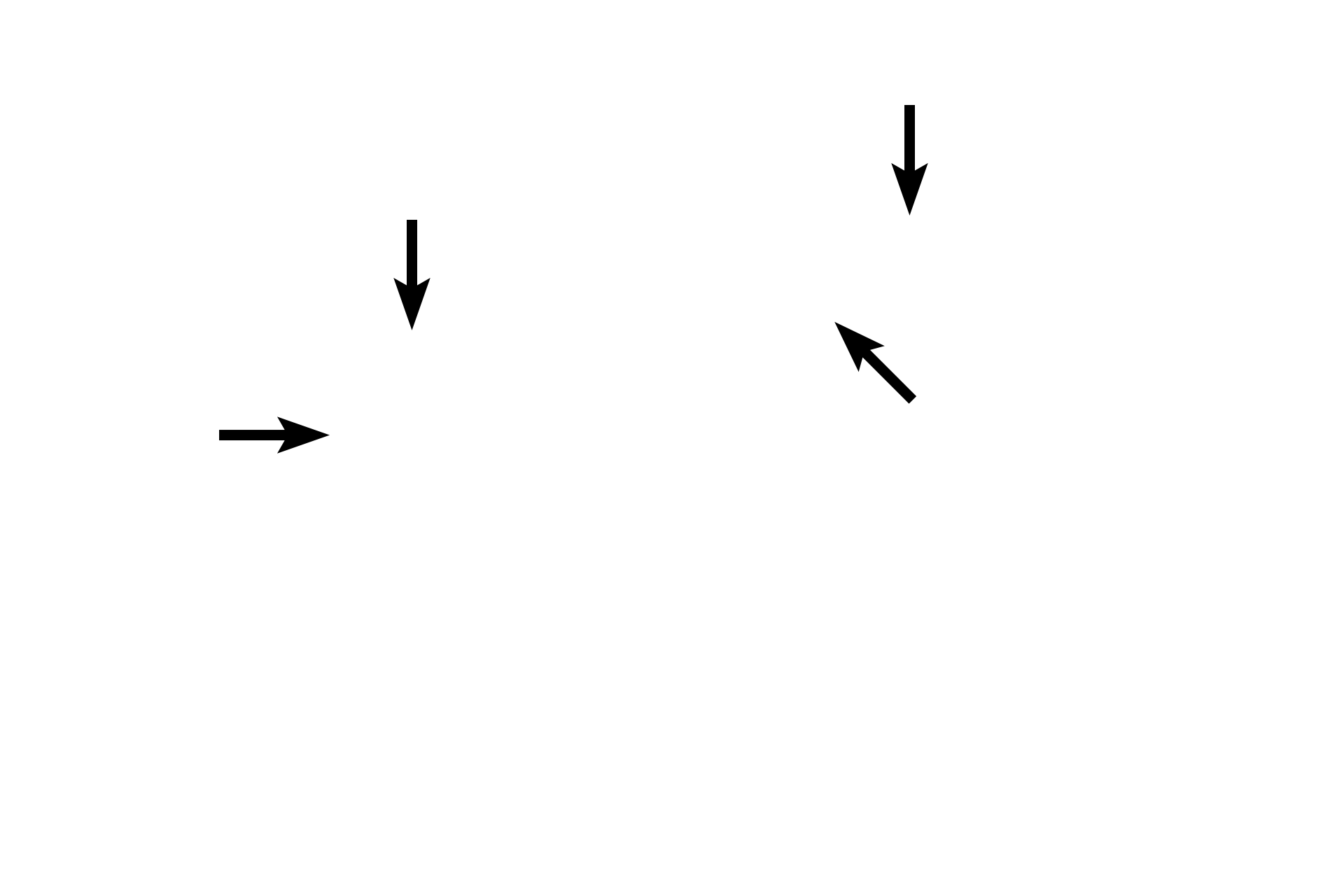
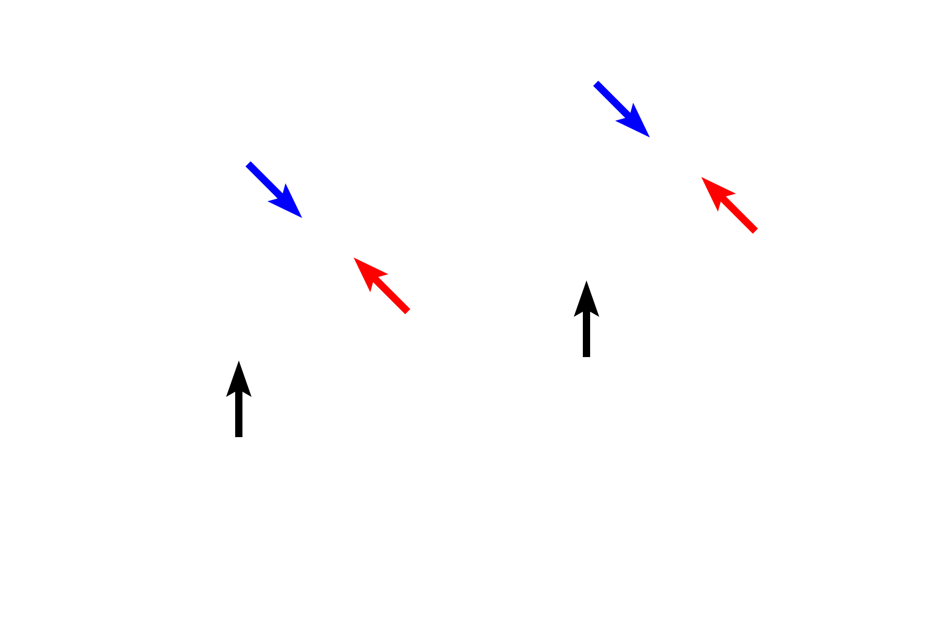
The mitotic spindle consists of two types of microtubules. Kinetochore microtubules (blue arrows) attach to chromatids, drawing them toward the spindle pole. Non-kinetochore microtubules are of two types. One set overlaps opposing microtubules to push the spindle poles away from the midline (red arrows); the second extends radially from the spindle apparatus (black arrows).