Stomach: fundus and body
This image of fundic glands demonstrates the cells of the glands, as well as the adjacent lamina propria and muscularis mucosae. 800x

Lumen of gland
This image of fundic glands demonstrates the cells of the glands, as well as the adjacent lamina propria and muscularis mucosae. 1000x

Muscularis mucosae
This image of fundic glands demonstrates the cells of the glands, as well as the adjacent lamina propria and muscularis mucosae. 1000x
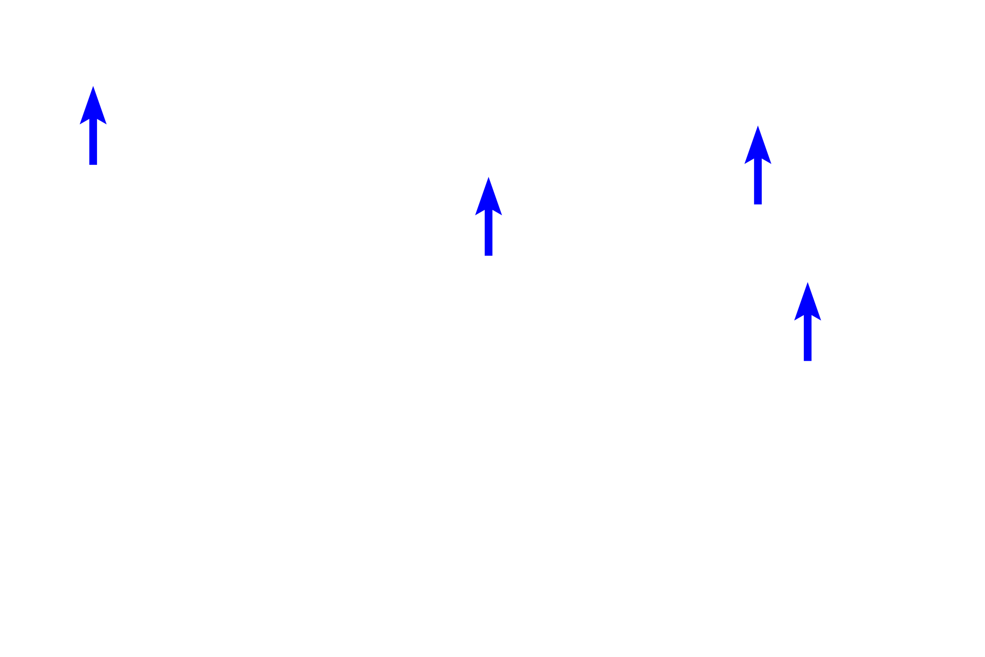
 Chief cells (black arrows) secrete the inactive form of the proteolytic enzyme pepsin, called pepsinogen. Pepsinogen is activated to pepsin in the lumen of the stomach. Nuclei and RER are located in the darker-staining bases of chief cells; the apical regions, appearing foamy, contain zymogen (secretory) granules (blue arrows) for release into the lumen of the gland.
">
Chief cells >
Chief cells (black arrows) secrete the inactive form of the proteolytic enzyme pepsin, called pepsinogen. Pepsinogen is activated to pepsin in the lumen of the stomach. Nuclei and RER are located in the darker-staining bases of chief cells; the apical regions, appearing foamy, contain zymogen (secretory) granules (blue arrows) for release into the lumen of the gland.

Enteroendocrine >
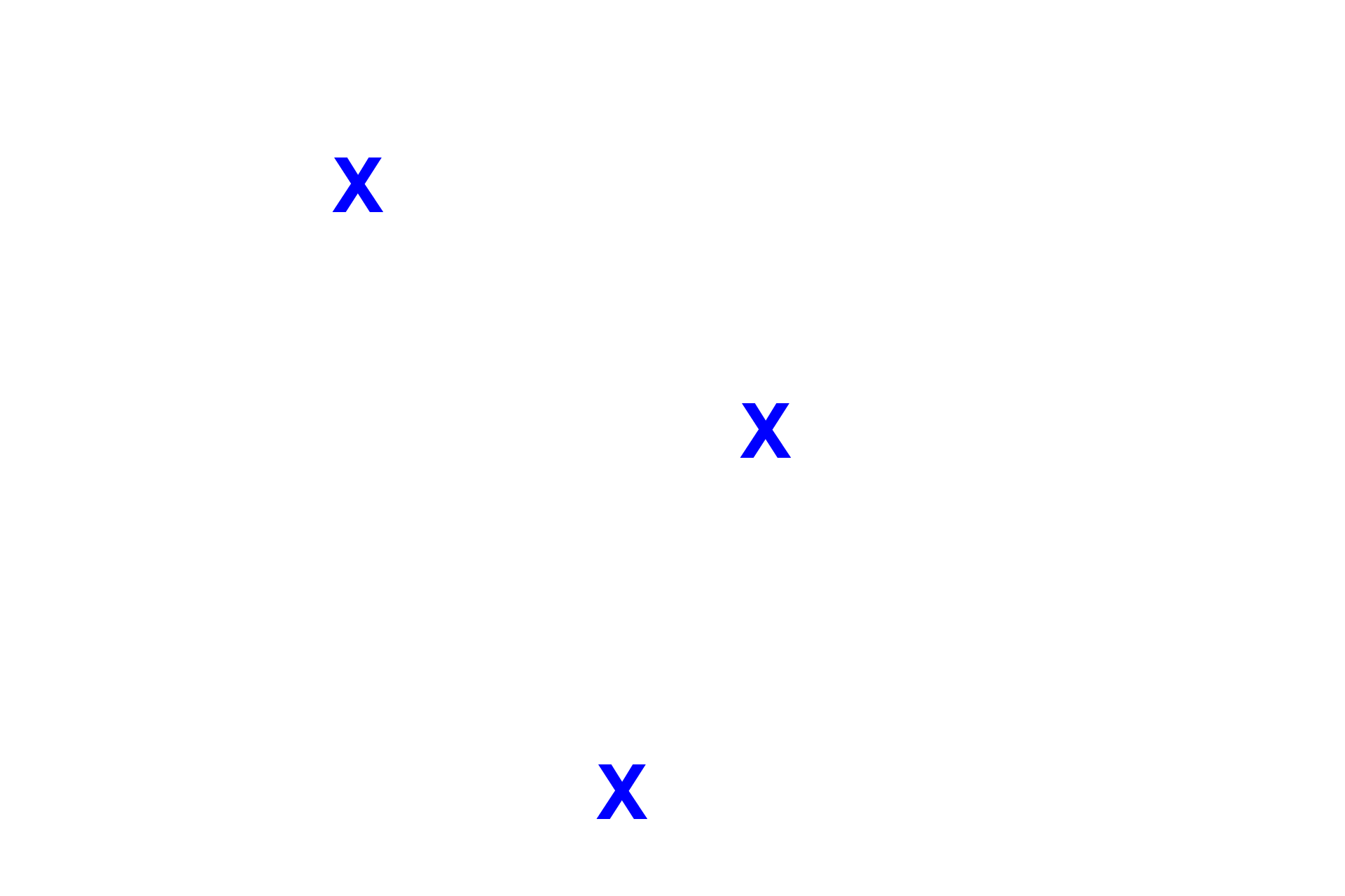
Enteroendocrine cells represent at least 12 different cell types, each secreting a specific hormone. These cells lie at the periphery of each gland, because they release their hormones into the lamina propria, where they enter blood vessels (endocrine) or influence adjacent exocrine cells (paracrine). Enteroendocrine cells are part of the diffuse neuroendocrine system (DNES).

Lamina propria
The lamina propria contains fibroblasts that maintain the connective tissue, as well as cells associated with the immune system.
 PREVIOUS