Adrenal gland
A higher magnification image of the adrenal shows both the cortex, surrounded by the dense connective tissue capsule, and the medulla. The adrenal cortex, source of the adrenal steroid hormones, is divided into a zona glomerulosa beneath the capsule, followed by the zona fasciculata and zona reticularis. 200x

Capsule
A higher magnification image of the adrenal shows both the cortex, surrounded by the dense connective tissue capsule, and the medulla. The adrenal cortex, source of the adrenal steroid hormones, is divided into a zona glomerulosa beneath the capsule, followed by the zona fasciculata and zona reticularis. 200x

Cortex >
The cortex of the adrenal gland is the steroid-secreting subdivision of the adrenal. As such, it is composed of cells filled with lipid droplets containing the precursors of steroid hormones. The lipid droplets give the cells a frothy appearance. Cortical cells also have abundant smooth endoplasmic reticulum.

- Zona glomerulosa >
The zona glomerulosa, the outermost and thinnest layer of the cortex, is composed of steroid-secreting cells that are clustered into spherical shapes. (“Glomus” means “ball” in Latin.) Zona glomerulosa cells secrete the mineralocorticoids.

- Zona fasciculata >
Zona fasciculata is the middle and thickest layer of the adrenal cortex; its cells are the most vacuolated in the cortex and are arranged in fascicles separated by wide-diameter, fenestrated capillaries. Its steroid-producing cells secrete glucocorticoids and androgens and have mitochondria with tubular cristae.

- Zona reticularis >
Zona reticularis is the innermost portion of the cortex, forming a meshwork of cells surrounded by wide-bore, fenestrated capillaries. Zona reticularis cells possess mitochondria with tubular cristae and abundant SER; these cells are less vacuolated and more eosinophilic than those in zona fasciculata. Glucocorticoids and androgens are also secreted by this layer.

Medulla >
The medulla forms the core of the adrenal gland. Cells of the medulla, derived from neural crest cells, are modified adrenergic neurons without axons or dendrites. These cells function as a sympathetic ganglion and are innervated by preganglionic sympathetic fibers. The catecholamines, norepinephrine and epinephrine, are released from the medulla.

Vasculature >
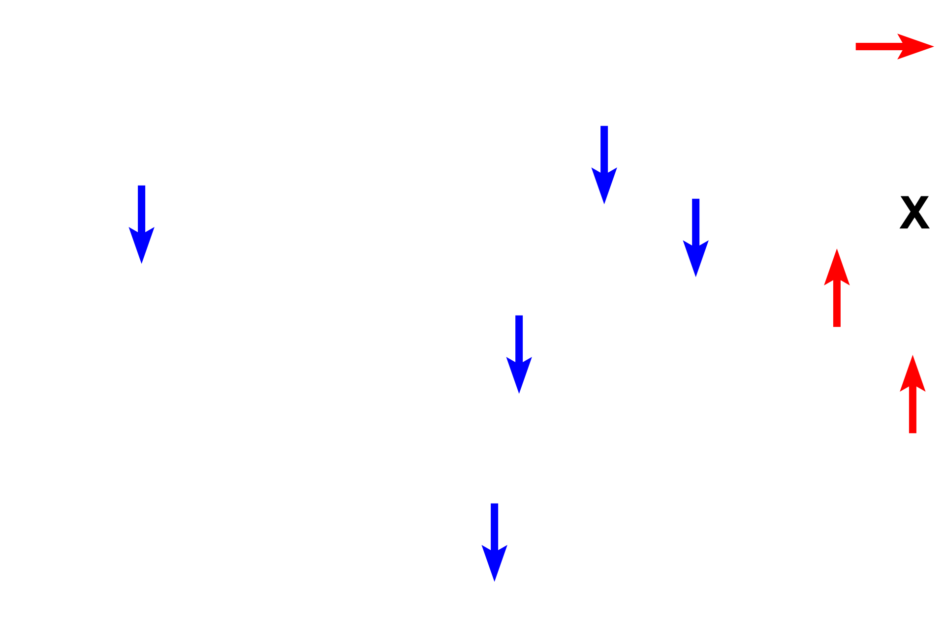
Arterioles in the capsule supply wide-bore fenestrated capillaries (blue arrows) with blood that percolates through cortical zones to enter medullary capillaries. Additional arterioles from the capsule directly supply the medullary capillaries (red arrows). Both capillary beds drain into larger veins (X) in the medulla, which anastomose to form the single medullary vein that exits from the organ.
 PREVIOUS