
Ovary: atretic follicles
Atretic follicles are the degenerating remains of follicles. Approximately two million primordial follicles, containing primary oocytes, are present at birth, but only 400-450 secondary oocytes are ovulated from Graafian follicles during the reproductive life of a woman; the remaining follicles undergo atresia. The process of atresia can occur at all stages of follicular development. 100x
Frame A >
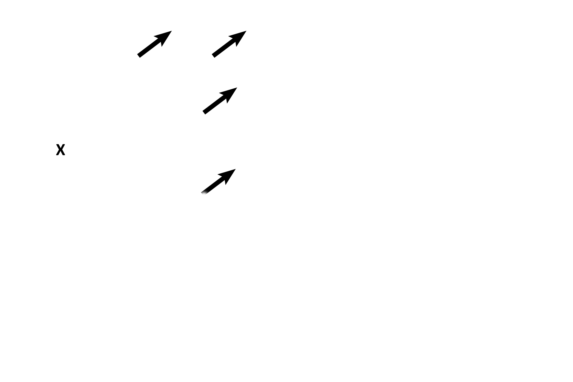
Two different types of atretic follicles are seen. The antrum of an atretic secondary follicle (X) is visible; note the granulosal cells sloughed into the antrum. Each atretic follicle labeled with an arrow is composed only of its zona pellucida; the remainder of each follicle has degenerated.
Frame B >
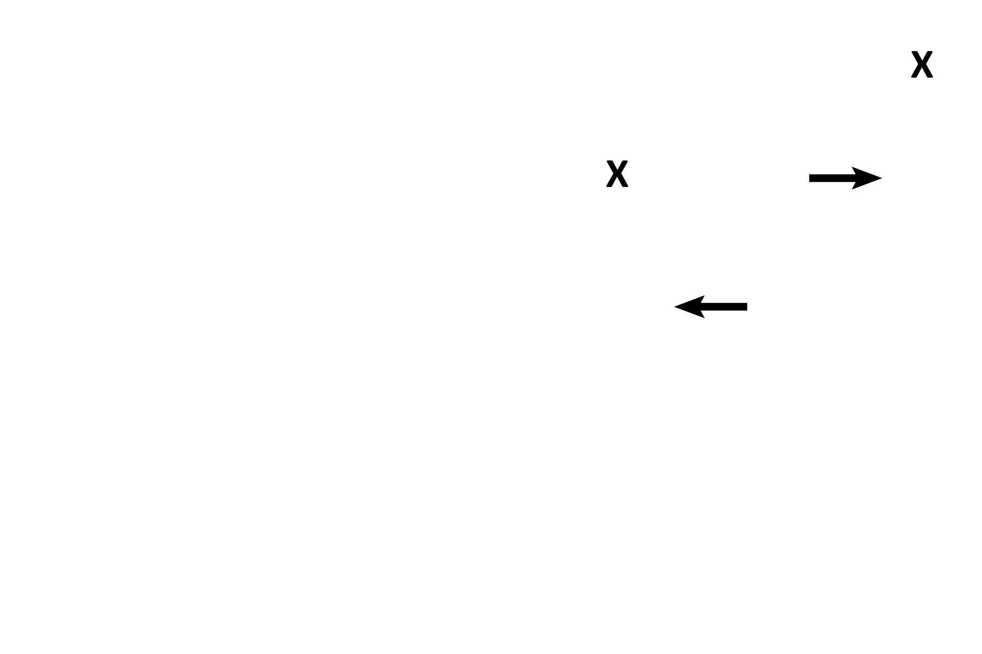
Portions of the atretic remains of two secondary follicles are seen; each antral space is labeled with an “X.” Each atretic follicle shows granulosal cells sloughing into the antrum and pulling away from the basement membrane (arrows).

Frame C >
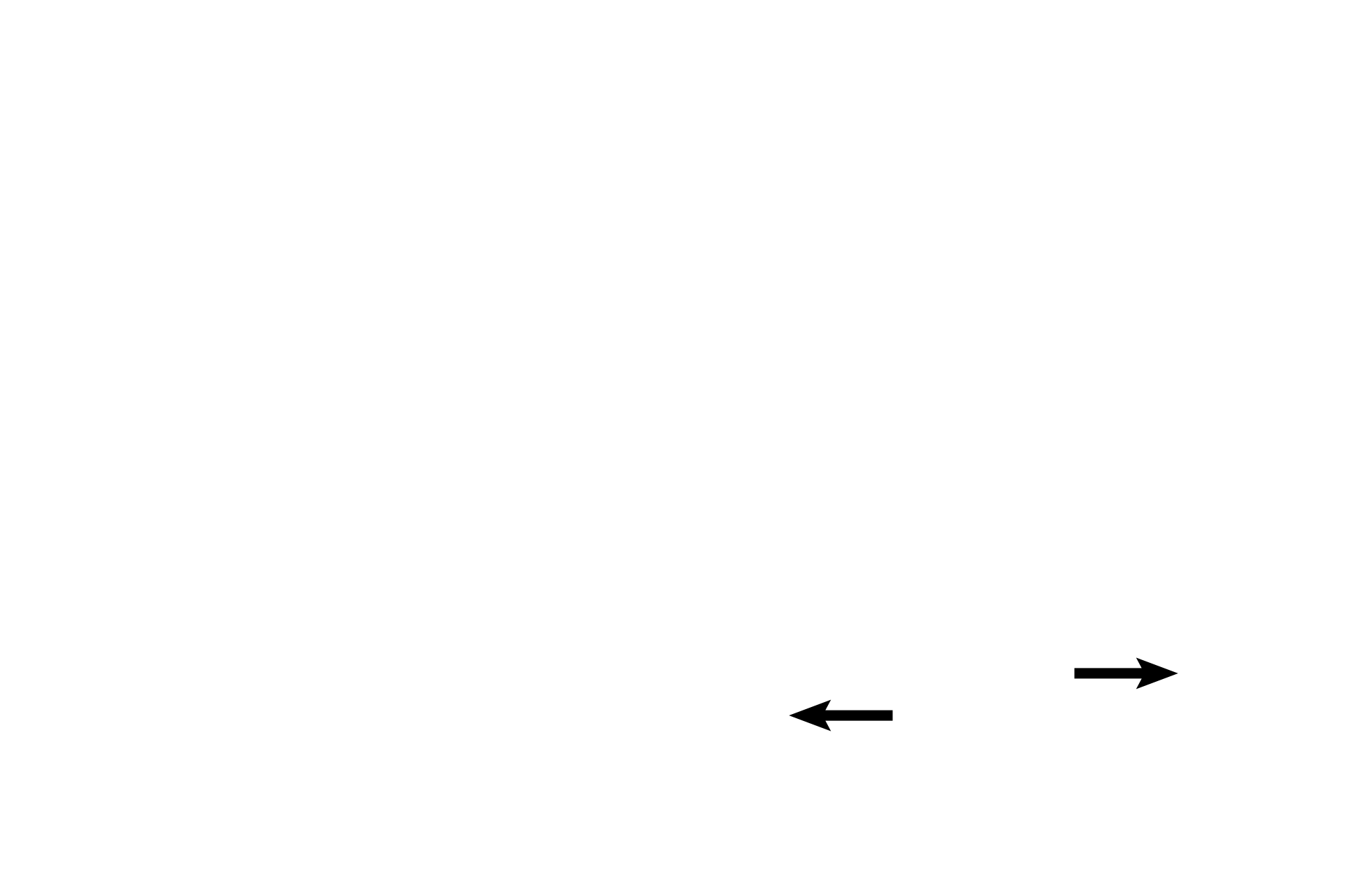
Three atretic follicles can be seen. An atretic secondary follicle (X) displays granulosa cells sloughing into the antrum. On the left (green arrow), the granulosal cells have disappeared, leaving the remnants of the oocyte and its zona pellucida denuded. The black arrow indicates the zona pellucida of another follicle; the remainder of this follicle has degenerated.
Frame D >
Both atretic follicles here are composed of the hypertrophied basement membranes that originally surrounded the follicular cells. As the cells of the follicles degenerate, the basement membrane hypertrophies and folds up on itself, resembling a crumpled rubber band. The atretic follicle on the left is older.