
Bone remodeling
These images through the diaphysis of a long bone demonstrate the multiple processes occurring during bone growth and development. The image on the right is later in time during the remodeling process than is the image on the left. 400x, 200x
Marrow space
These images, through the diaphysis of a long bone, demonstrate the multiple processes occurring during bone growth and development. The image on the right is later in time during the remodeling process than is the image on the left. 400x, 200x

Periosteum
These images, through the diaphysis of a long bone, demonstrate the multiple processes occurring during bone growth and development. The image on the right is later in time during the remodeling process than is the image on the left. 400x, 200x
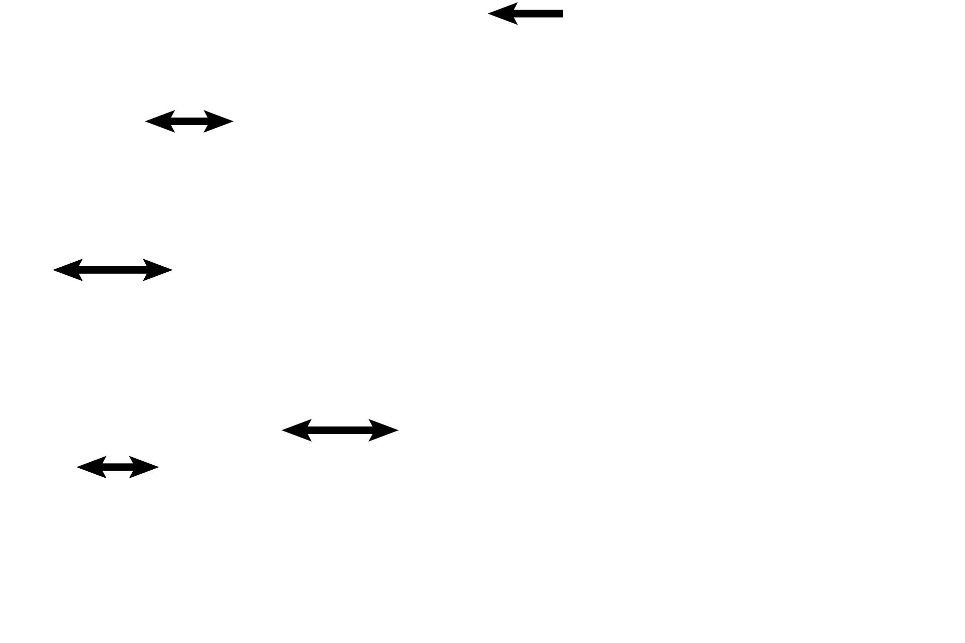
Circumferential lamellae >
Inner (1) and outer (2) circumferential lamellae can be seen on either surface of the bone, indicating an increase in the diameter of the bone. This increased diameter provides additional support as an individual grows in mass. Circumferential lamellae are deposited by intramembranous bone formation by the endosteal and periosteal membranes.
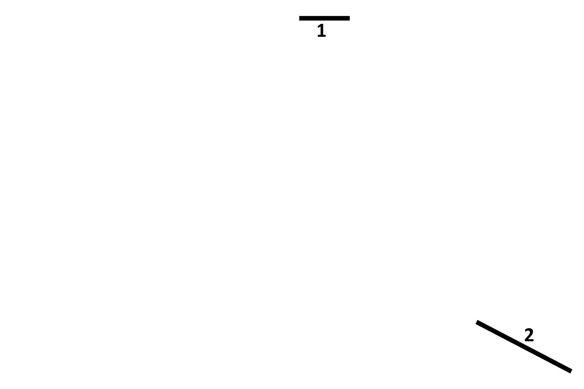
Calcified cartilage >
Islands of acellular tissue are present between the two stacks of circumferential lamellae and in the spongy bone. These areas represent the calcified cartilage remnants from the epiphyseal plate. The presence of this calcified cartilage indicates that this central area was produced by endochondral ossification.
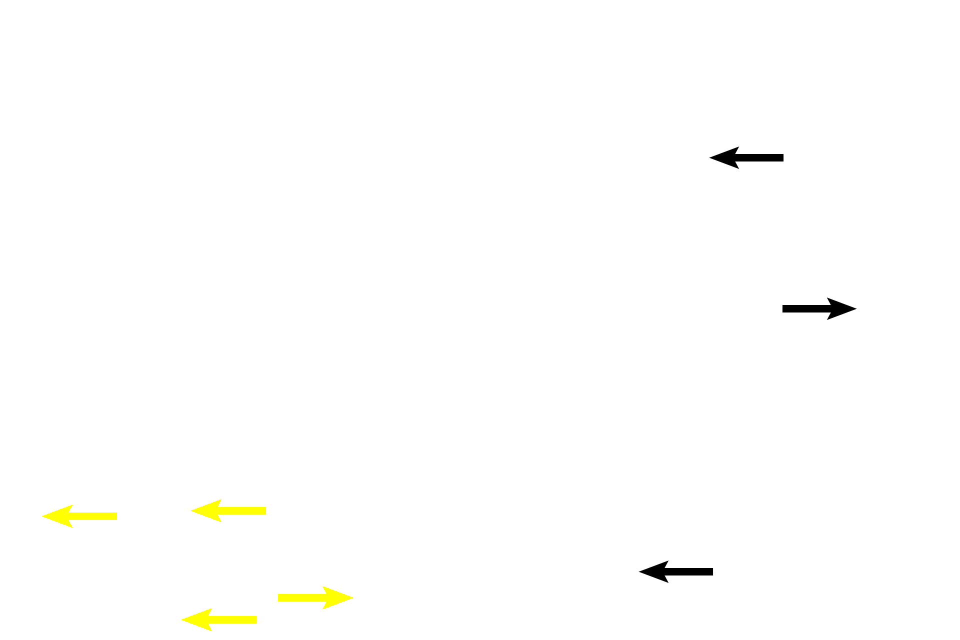
Woven bone >
Immediately surrounding the calcified cartilage are small areas of woven bone, remnants of the zone of ossification. Because this bone is located in a space previously occupied by cartilage, this bone was formed by endochondral ossification. Both the calcified cartilage and the woven bone will be resorbed in time, to be replaced with lamellar bone.

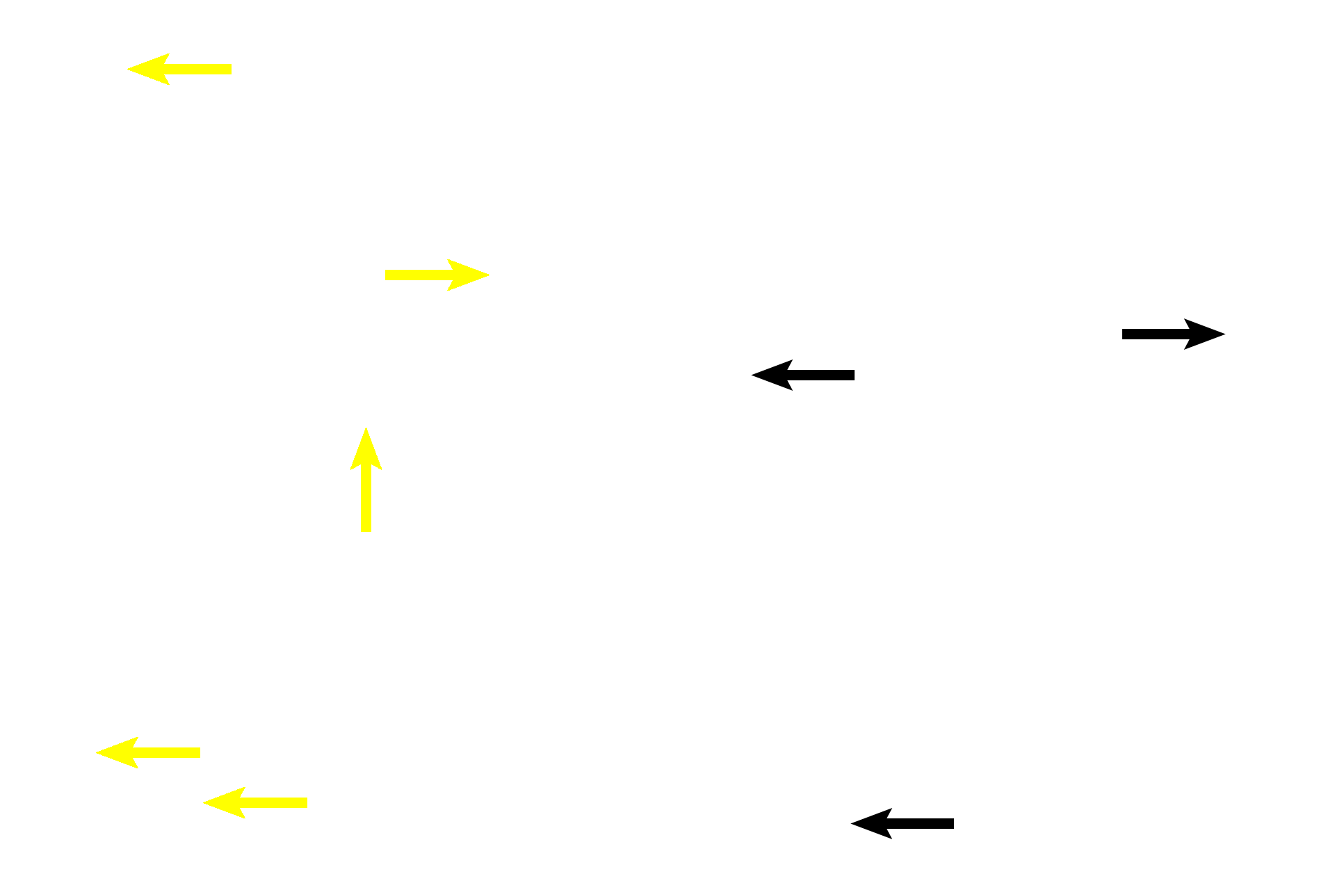
New lamellae >
Lamellar deposition has begun around the periphery of several spaces, which were created by death of chondrocytes during endochondral ossification. Continued deposition will create osteons, converting this spongy woven bone to the compact bone seen in the center of the image on the right.
Osteons
Lamellar deposition has begun around the periphery of several spaces, which were created by death of chondrocytes during endochondral ossification. Continued deposition will create osteons, converting this spongy woven bone to the compact bone seen in the center of the image on the right.
Cement lines >
Cement lines separate bone deposited at different time periods.