Cortex: Convoluted portion
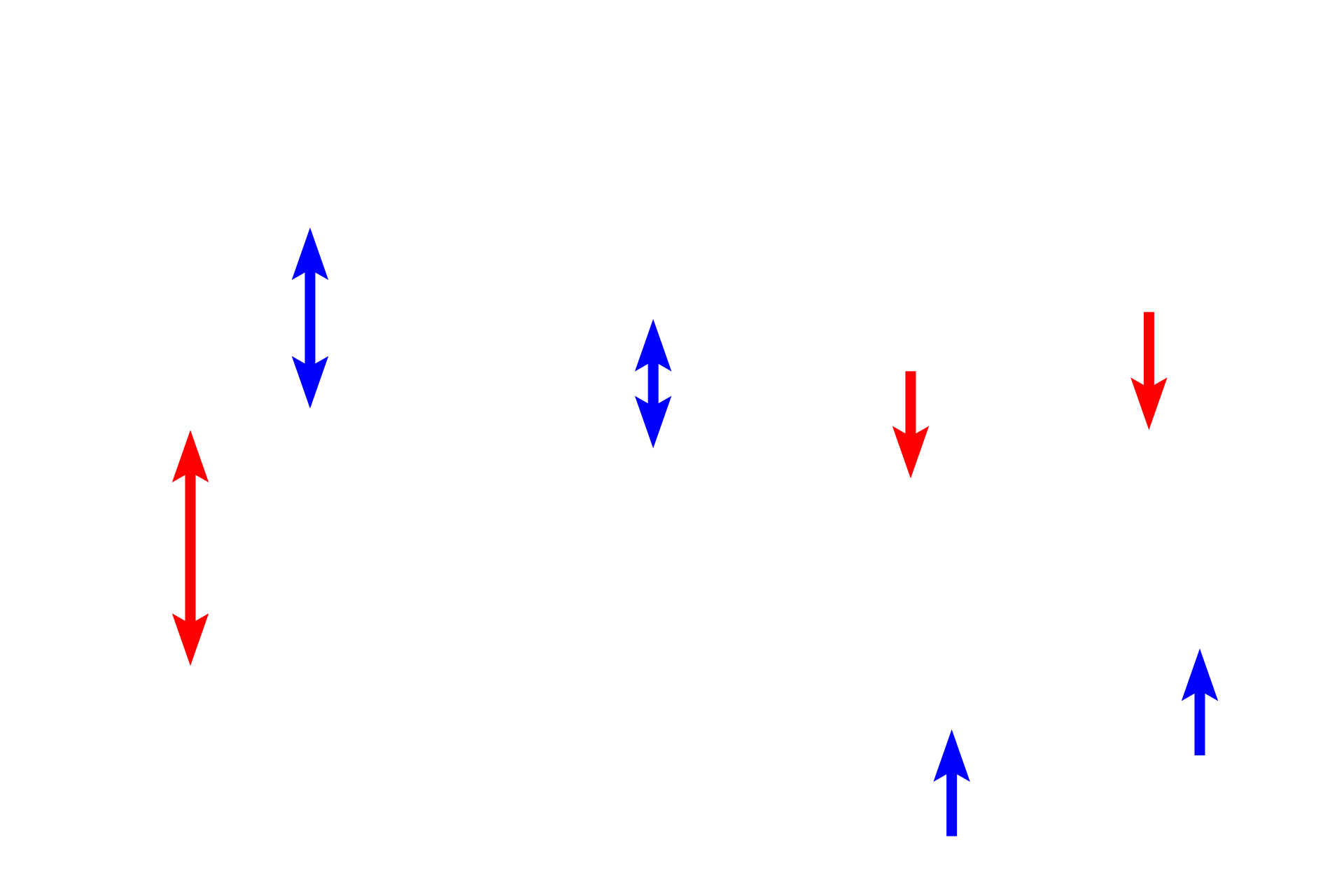
The histological appearances of proximal and distal tubules are contrasted in this image. 600x

Proximal tubules >
Proximal tubules are larger in diameter and have a taller, simple cuboidal epithelium than do distal tubules. The presence of a prominent brush border (composed of microvilli) also helps to distinguish the proximal tubules. At the light microscopic level, convoluted and straight portions of proximal tubules are similar but can be distinguished by their location.

- Brush border
Proximal tubules are larger in diameter and have a taller, simple cuboidal epithelium than do distal tubules. The presence of a prominent brush border (composed of microvilli) also helps to distinguish the proximal tubules. At the light microscopic level, convoluted and straight portions of proximal tubules are similar but can be distinguished by their location.

Distal tubules >
Distal tubules, ascending thick limb (blue arrows) and distal convoluted tubules (red arrows) are also lined by a simple cuboidal epithelium. The epithelium lacks a brush border and varies in height depending on the tubule type, with the convoluted tubule have a shorter epithelium. Additionally, epithelial nuclei are often irregularly spaced around and frequently bulge into the lumen, particularly when the epithelium is short.

Peritubular capillaries >
Peritubular capillaries surround the tubules of the nephron and are supplied by the efferent arteriole.
 PREVIOUS