
Endochondral ossification
A cross section though a long bone shows the periosteal collar encasing spicules that have a blue central core and a periphery of red tissue. Such staining qualities indicate that both cartilage and bone are present and that, therefore, either the zone of ossification or the zone of resorption or both are represented in this section. 40x
Periosteal collar >
A periosteal collar of woven bone forms the bony periphery. This bone, laid down by intramembranous bone formation, forms the diaphysis of this fetal, long bone. The periosteal collar was formed and is covered on its outer surface by a periosteum.

Periosteum
A periosteal collar of woven bone forms the bony periphery. This bone, laid down by intramembranous bone formation, forms the diaphysis of this fetal, long bone. The periosteal collar was formed and is covered on its outer surface by a periosteum.
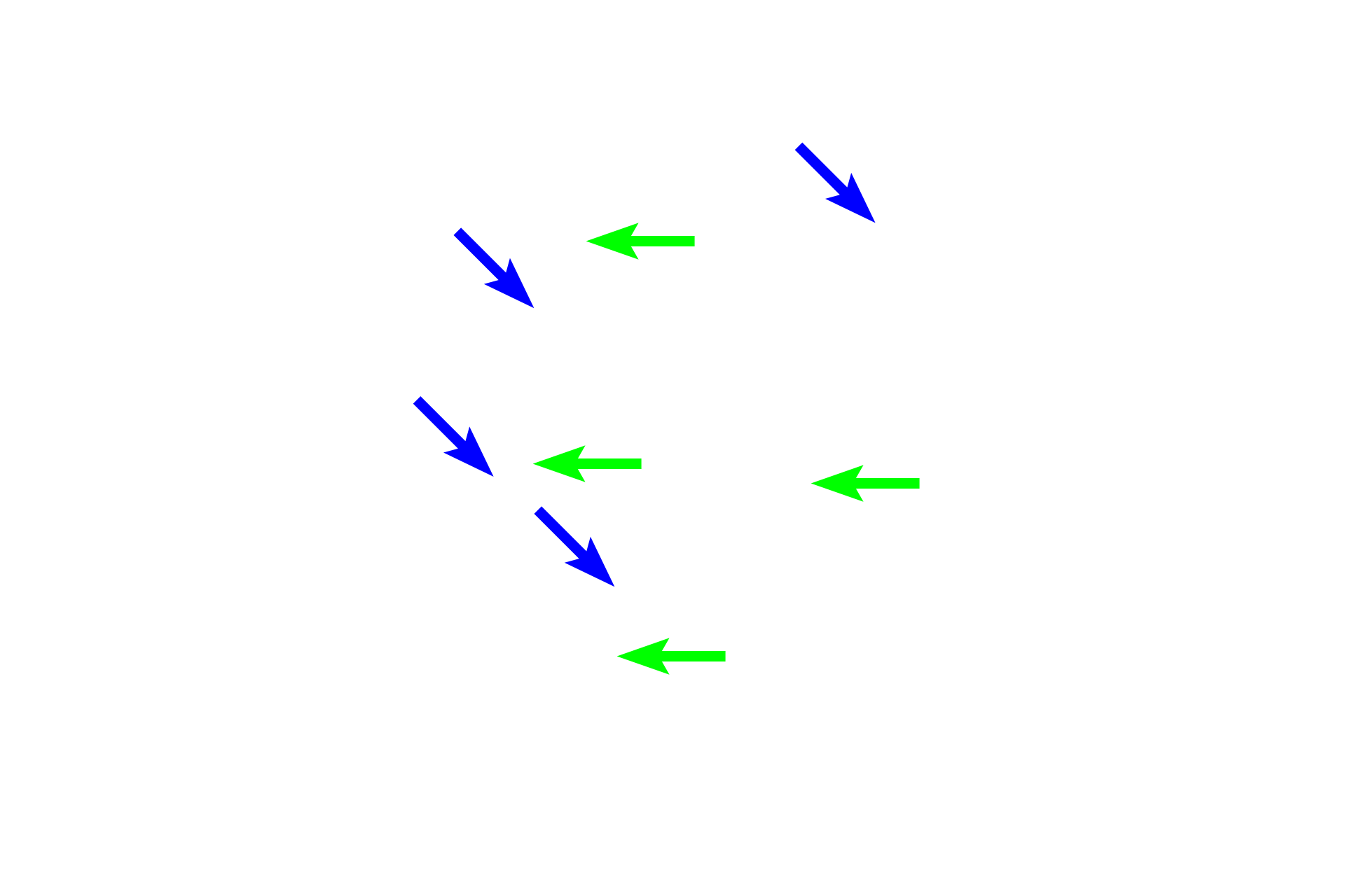
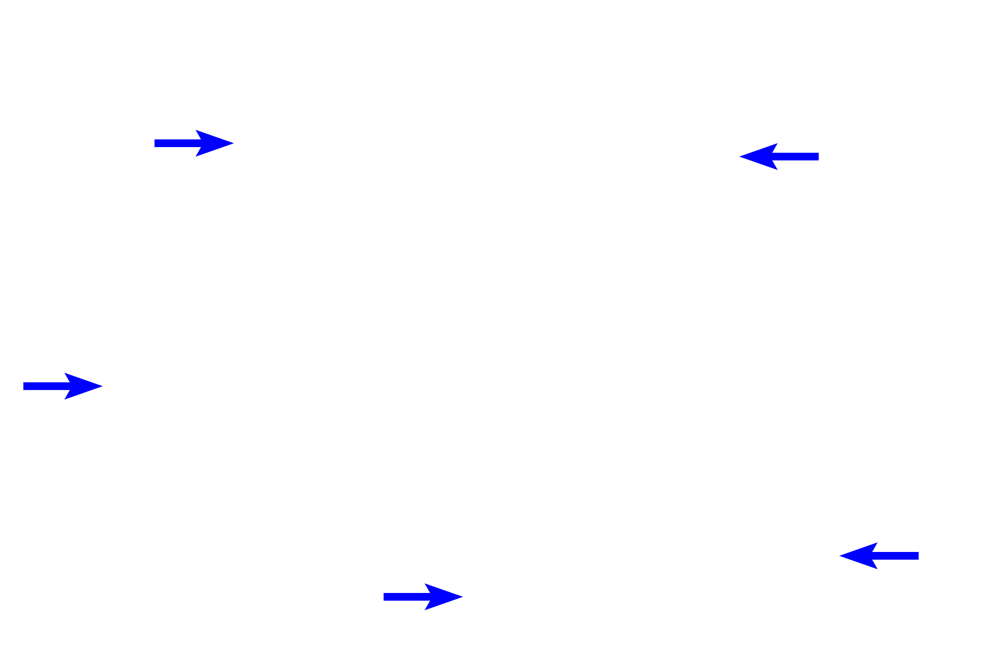
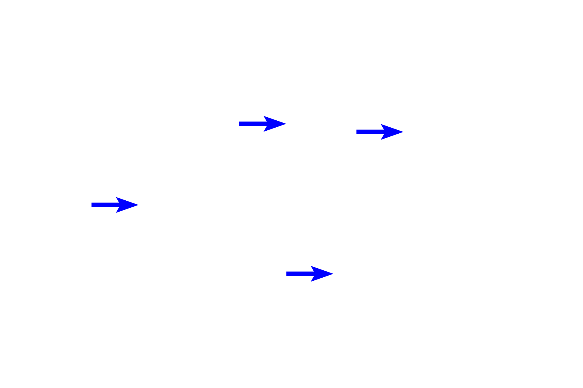
Spicules >
Spicules in this section consist of both calcified cartilage (green arrows) and bone (blue arrows). They are eventually resorbed by osteoclasts in the zone of resorption.
Red marrow >
Hemopoietic red bone marrow occupies the spaces between the spicules.